

Cross-organ sensitization of thoracic spinal neurons receiving noxious cardiac input in rats with gastroesophageal reflux
- PMID: 20378832
- PMCID: PMC3774335
- DOI: 10.1152/ajpgi.00312.2009
Cross-organ sensitization of thoracic spinal neurons receiving noxious cardiac input in rats with gastroesophageal reflux
Abstract
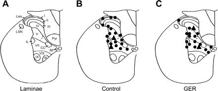
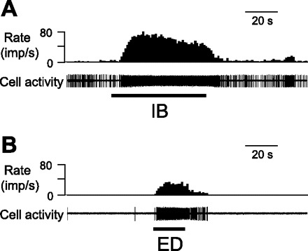
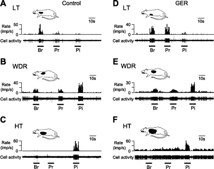
Gastroesophageal reflux (GER) frequently triggers or worsens cardiac pain or symptoms in patients with coronary heart disease. This study aimed to determine whether GER enhances the activity of upper thoracic spinal neurons receiving noxious cardiac input. Gastric fundus and pyloric ligations as well as a longitudinal myelotomy at the gastroesophageal junction induced acute GER in pentobarbital-anesthetized, paralyzed, and ventilated male Sprague-Dawley rats. Manual manipulations of the stomach and lower esophagus were used as surgical controls in another group. At 4-9 h after GER surgery, extracellular potentials of single neurons were recorded from the T3 spinal segment. Intrapericardial bradykinin (IB) (10 microg/ml, 0.2 ml, 1 min) injections were used to activate cardiac nociceptors, and esophageal distensions were used to activate esophageal afferent fibers. Significantly more spinal neurons in the GER group responded to IB compared with the control group (69.1 vs. 38%, P < 0.01). The proportion of IB-responsive neurons in the superficial laminae of GER animals was significantly different from those in deeper layers (1/8 vs. 46/60, P < 0.01); no difference was found in control animals (7/25 vs. 20/46, P > 0.05). Excitatory responses of spinal neurons to IB in the GER group were greater than in the control group [32.4 +/- 3.5 impulses (imp)/s vs. 13.3 +/- 2.3 imp/s, P < 0.01]. Forty-five of 47 (95.7%) neurons responded to cardiac input and ED, which was higher than the control group (61.5%, P < 0.01). These results indicate that acute GER enhanced the excitatory responses of thoracic spinal neurons in deeper laminae of the dorsal horn to noxious cardiac stimulus.
Figures

Similar articles
-
Neuromodulation of thoracic intraspinal visceroreceptive transmission by electrical stimulation of spinal dorsal column and somatic afferents in rats.J Pain. 2008 Jan;9(1):71-8. doi: 10.1016/j.jpain.2007.08.007. Epub 2007 Nov 5. J Pain. 2008. PMID: 17974489 Free PMC article.
-
Gastrocardiac afferent convergence in upper thoracic spinal neurons: a central mechanism of postprandial angina pectoris.J Pain. 2007 Jun;8(6):522-9. doi: 10.1016/j.jpain.2007.02.428. Epub 2007 Apr 16. J Pain. 2007. PMID: 17434802
-
Responses and afferent pathways of superficial and deeper c(1)-c(2) spinal cells to intrapericardial algogenic chemicals in rats.J Neurophysiol. 2001 Apr;85(4):1522-32. doi: 10.1152/jn.2001.85.4.1522. J Neurophysiol. 2001. PMID: 11287476
-
Gastroesophageal reflux in childhood.Curr Probl Surg. 1996 Jan;33(1):1-70. Curr Probl Surg. 1996. PMID: 8536488 Review.
-
[Neurobiology of visceral pain].Schmerz. 2014 Jun;28(3):233-51. doi: 10.1007/s00482-014-1402-x. Schmerz. 2014. PMID: 24903037 Review. German.
Cited by
-
Studying Cardiac Neural Network Dynamics: Challenges and Opportunities for Scientific Computing.Front Physiol. 2022 Apr 29;13:835761. doi: 10.3389/fphys.2022.835761. eCollection 2022. Front Physiol. 2022. PMID: 35574437 Free PMC article. Review.
-
Chronic Prostatitis Induces Bladder Hypersensitivity and Sensitizes Bladder Afferents in the Mouse.J Urol. 2016 Sep;196(3):892-901. doi: 10.1016/j.juro.2016.03.077. Epub 2016 Mar 17. J Urol. 2016. PMID: 26997315 Free PMC article.
-
Co-occurrence of pain syndromes.J Neural Transm (Vienna). 2020 Apr;127(4):625-646. doi: 10.1007/s00702-019-02107-8. Epub 2019 Nov 29. J Neural Transm (Vienna). 2020. PMID: 31784821 Review.
References
-
- Albutaihi IA, DeJongste MJ, Ter Horst GJ. An integrated study of heart pain and behavior in freely moving rats (using fos as a marker for neuronal activation). Neurosignals 13: 207–226, 2004 - PubMed
-
- Banerjee B, Medda BK, Lazarova Z, Bansal N, Shaker R, Sengupta JN. Effect of reflux-induced inflammation on transient receptor potential vanilloid one (TRPV1) expression in primary sensory neurons innervating the oesophagus of rats. Neurogastroenterol Motil 19: 681–691, 2007 - PubMed
-
- Bennett JR, Atkinson M. The differentiation between oesophageal and cardiac pain. Lancet 2: 1123–1127, 1966 - PubMed
-
- Brook RA, Wahlqvist P, Kleinman NL, Wallander MA, Campbell SM, Smeeding JE. Cost of gastro-oesophageal reflux disease to the employer: a perspective from the United States. Aliment Pharmacol Ther 26: 889–898, 2007 - PubMed
-
- Castell DO. Chest pain of undetermined origin: overview of pathophysiology. Am J Med 92: 2S–4S, 1992 - PubMed
Publication types
MeSH terms
Grants and funding
LinkOut - more resources
Full Text Sources
Medical