. 2010 May 28;106(10):1603-12.
doi: 10.1161/CIRCRESAHA.110.216804. Epub 2010 Apr 8.

Isoform-selective physical coupling of TRPC3 channels to IP3 receptors in smooth muscle cells regulates arterial contractility

Affiliations

Isoform-selective physical coupling of TRPC3 channels to IP3 receptors in smooth muscle cells regulates arterial contractility

Adebowale Adebiyi et al. Circ Res. .

Abstract

Rationale: Inositol 1,4,5-trisphosphate (IP(3))-induced vasoconstriction can occur independently of intracellular Ca(2+) release and via IP(3) receptor (IP(3)R) and canonical transient receptor potential (TRPC) channel activation, but functional signaling mechanisms mediating this effect are unclear.

Objectives: Study mechanisms by which IP(3)Rs stimulate TRPC channels in myocytes of resistance-size cerebral arteries.

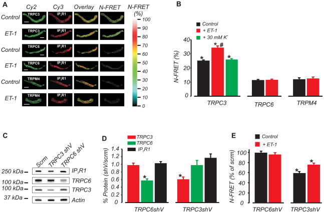
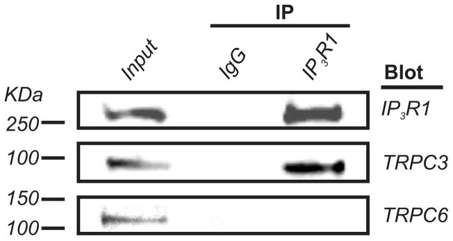
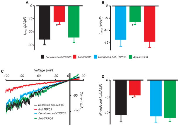
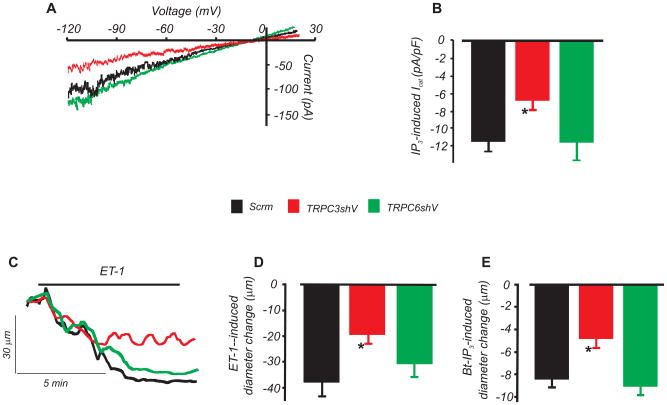
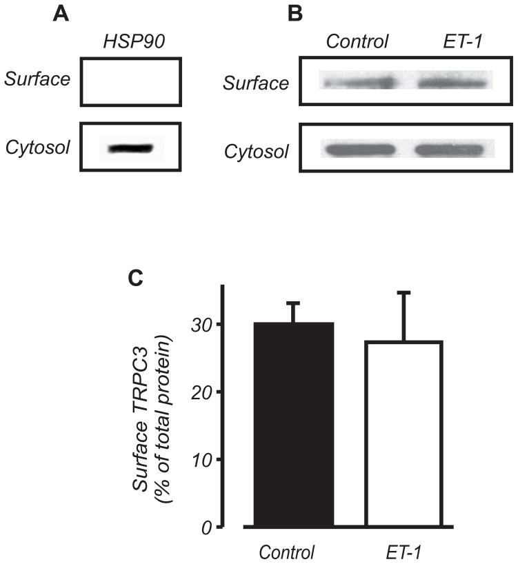
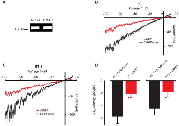
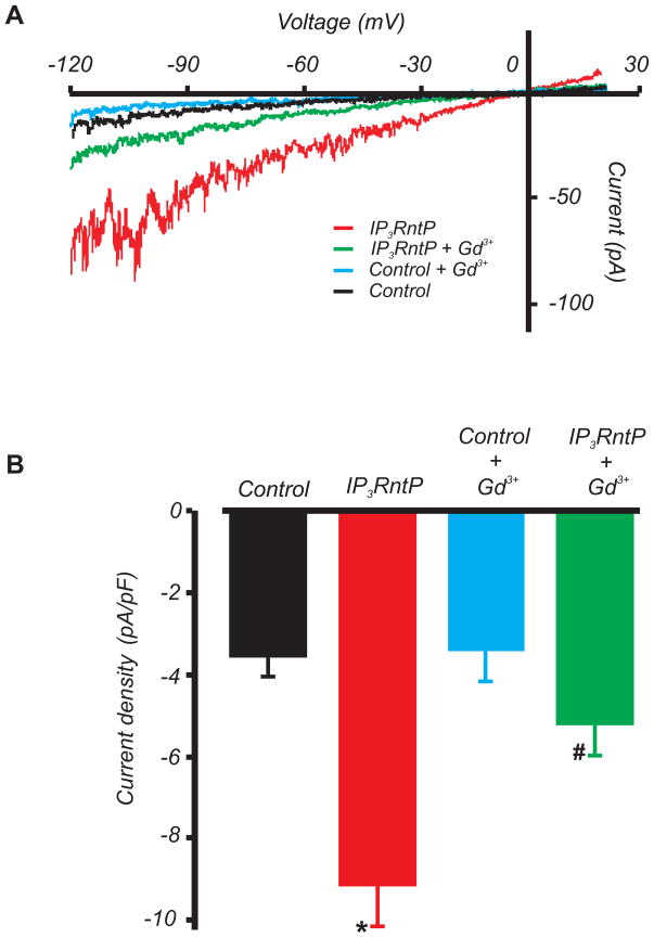
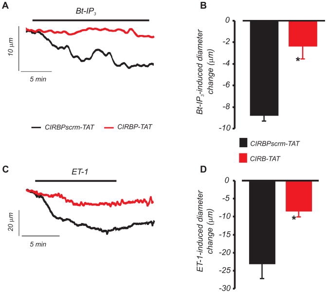
Methods and results: Immunofluorescence resonance energy transfer (immuno-FRET) microscopy using isoform-selective antibodies indicated that endogenous type 1 IP(3)Rs (IP(3)R1) are in close spatial proximity to TRPC3, but distant from TRPC6 or TRPM4 channels in arterial myocytes. Endothelin-1 (ET-1), a phospholipase C-coupled receptor agonist, elevated immuno-FRET between IP(3)R1 and TRPC3, but not between IP(3)R1 and TRPC6 or TRPM4. TRPC3, but not TRPC6, coimmunoprecipitated with IP(3)R1. TRPC3 and TRPC6 antibodies selectively inhibited recombinant channels, but only the TRPC3 antibody blocked IP(3)-induced nonselective cation current (I(Cat)) in myocytes. TRPC3 knockdown attenuated immuno-FRET between IP(3)R1 and TRPC3, IP(3)-induced I(Cat) activation, and ET-1 and IP(3)-induced vasoconstriction, whereas TRPC6 channel knockdown had no effect. ET-1 did not alter total or plasma membrane-localized TRPC3, as determined using surface biotinylation. RT-PCR demonstrated that C-terminal calmodulin and IP(3)R binding (CIRB) domains are present in myocyte TRPC3 and TRPC6 channels. A peptide corresponding to the IP(3)R N-terminal region that can interact with TRPC channels activated I(Cat). A TRPC3 CIRB domain peptide attenuated IP(3)- and ET-1-induced I(Cat) activation and vasoconstriction.

Conclusions: IP(3) stimulates direct coupling between IP(3)R1 and membrane-resident TRPC3 channels in arterial myocytes, leading to I(Cat) activation and vasoconstriction. Close spatial proximity between IP(3)R1 and TRPC3 establishes this isoform-selective functional interaction.


Figures

Figure 1
Figure 1. TRPC3, but not TRPM4 or TRPC6, is in close spatial proximity to IP3R1 channels in arterial myocytes
A, Fluorescent images of individual Cy2 and Cy3 labels, pixel overlay, and N-FRET for indicated TRP channel primary antibody combinations. Scale bar=10 μm; B, Mean data illustrating control and ET-1 (100 nmol/L)-induced N-FRET generated by Cy3 bound IP3R1 and Cy2-bound TRPC3, TRPM4, or TRPC6 antibodies, and effects of ET-1 and 30 mmol/L K+. * P<0.05 compared with TRPC6 or TRPM4; # P<0.05 compared with IP3R1-TRPC3 in control. n for columns from left to right are=27, 11, 15, 20, 28, 17, and 13, respectively. C, Representative Western blot illustrating that TRPC3shV causes selective knockdown of TRPC3 and TRPC6shV induces selective knockdown of TRPC6. D, Mean data illustrating effects of TRPC3shV and TRPC6shV on TRPC3 (n=4 and 6), TRPC6 (n=4 and 6), and IP3R1 (n=4 each), respectively (* P<0.05). E, TRPC6 knockdown did not reduce the N-FRET signal between IP3R1 and TRPC6 in control (n=15) or ET-1 (n=10), whereas TRPC3 knockdown reduced the N-FRET signal between IP3R1 and TRPC3 in control (n=10) and ET-1 (n=15). * P<0.05 compared with scrm.
Figure 2
Figure 2. IP3R1 interacts with TRPC3 but not TRPC6 channels in cerebral arteries
IP3R1 monoclonal antibody co-immunoprecipitates IP3R1 (~270 kDa) and TRPC3 (~90 kDa), but not TRPC6 (~110 kDa) in cerebral arteries. Lysate supernatant (20 μg protein) was used as the input control. Mouse IgG was used instead of the IP3R1 monoclonal antibody as a negative co-IP control.
Figure 3
Figure 3. TRPC3 mediates IP3-induced ICat in arterial myocytes
A,B, Mean data for current density generated in HEK293 cells in response to transfection with recombinant TRPC3 or TRPC6 channels, and selective inhibition by TRPC3 and TRPC6 antibodies (2 μg/mL each). Whole cell currents were essentially absent in cells transfected with only GFP. n for columns from left to right are = 7, 7, 6, 6, 6, and 4, respectively. C, Exemplary recordings illustrating inhibition of IP3 (10 μmol/L in pipette solution)-induced ICat by TRPC3 antibody (2 μg/mL), but no effect of TRPC6 antibody (2 μg/mL), in arterial myocytes. D, Mean data for active and denatured TRPC3 (n=8 each) and TRPC6 antibody (n=7 each) in arterial myocytes. *P<0.05 vs. denatured control.
Figure 4
Figure 4. TRPC3 knockdown attenuates IP3-induced ICat activation and vasoconstriction whereas TRPC6 knockdown has no effect
A, Exemplary traces of IP3 (10 μmol/L)-induced ICat in arterial myocytes treated with scrm, TRPC3shV, or TRPC6shV. B, Mean ICat density (n: scrm, 6; TRPC3shV; 10, TRPC6shV, 8). C, Exemplary recordings of ET-1-induced vasoconstriction in pressurized (20 mmHg) arteries treated with scrm, TRPC3shV, or TRPC6shV. D and E, Mean data for ET-1- (300 pmol/L) and Bt-IP3 (10 nmol/L)-induced vasoconstriction (n=5, 4, and 4 for scrm, TRPC3shV, and TRPC6shV, respectively). * P<0.05 versus scrm). Baseline tone was similar in scrm- (12 ± 4%, n=5), TRPC3shV- (10 ± 1%, n=4), and TRPC6shV- (11 ± 2%, n=4) treated arteries (P>0.05 for each).
Figure 5
Figure 5. ET-1 does not increase surface expression of TRPC3 channels in cerebral arteries
A, Western blot illustrating that HSP90 is detected only in the non-biotinylated (cytosolic) fraction in cerebral arteries. B,C, Western blot and mean data (n=3) indicating that ET-1 (10 nmol/L) does not increase plasma membrane-localized TRPC3.
Figure 6
Figure 6. IP3R to TRPC physical coupling is essential for ET-1- and IP3-induced ICat activation in arterial myocytes
A, Cerebral artery myocyte TRPC3 and TRPC6 channels both contain CIRB domains, as determined using nested RT-PCR. B and C, Exemplary recordings illustrating attenuation of IP3 (10 μmol/L) and ET-1 (100 nmol/L)-induced ICat activation by CIRBP (1 μmol/L via pipette) in arterial myocytes. D, Mean IP3 and ET-1-induced change in ICat density in CIRBP or CIRBPscrm (1 μmol/L each) -treated myocytes (*P<0.05 vs. CIRBPscrm; n=9, 9, 10, and 8 for ET-1+CIRBPscrm, ET-1+CIRBP, IP3+CIRBPscrm, and IP3+CIRBP, respectively).
Figure 7
Figure 7. IP3RntP activates non-selective cation currents in arterial myocytes
A, Exemplary recordings illustrating activation of Gd3+-sensitive ICat by IP3RntP (50 μmol/L via pipette). B, Mean ICat density. *P<0.05 vs. control; #P<0.05 vs. IP3RntP (n=5 and 6 for control and IP3RntP respectively).
Figure 8
Figure 8. IP3R to TRPC physical coupling contributes to ET-1 and IP3-induced vasoconstriction
A, B, CIRBP-TAT (3 μmol/L, 20 min application) attenuates Bt-IP3 (1 nmol/L, n=4)-induced vasoconstriction in pressurized (60 mmHg) cerebral arteries. C, D, CIRBP-TAT (3 μmol/L, 20 min application) attenuates ET-1 (1 nmol/L, n=7)-induced vasoconstriction (*P<0.05 vs. CIRBPscrm-TAT). When applied alone, CIRBP-TAT and CIRBPscrm-TAT similarly reduced mean baseline diameter by ~15 and 13 μm, respectively, consistent with a previous report for the TAT sequence.

References

    1. Berridge MJ. Inositol trisphosphate and calcium signalling. Nature. 1993;361:315–325. - PubMed
    1. Davis MJ, Hill MA. Signaling mechanisms underlying the vascular myogenic response. Physiol Rev. 1999;79:387–423. - PubMed
    1. Sanders KM. Invited review: mechanisms of calcium handling in smooth muscles. J Appl Physiol. 2001;91:1438–1449. - PubMed
    1. Xi Q, Adebiyi A, Zhao G, Chapman KE, Waters CM, Hassid A, Jaggar JH. IP3 constricts cerebral arteries via IP3 receptor-mediated TRPC3 channel activation and independently of sarcoplasmic reticulum Ca2+ release. Circ Res. 2008;102:1118–1126. - PMC - PubMed
    1. Zhao G, Adebiyi A, Blaskova E, Xi Q, Jaggar JH. Type 1 inositol 1,4,5-trisphosphate receptors mediate UTP-induced cation currents, Ca2+ signals, and vasoconstriction in cerebral arteries. Am J Physiol Cell Physiol. 2008;295:C1376–C1384. - PMC - PubMed

Publication types

MeSH terms

Substances

LinkOut - more resources