. 2010 May 28;106(10):1570-81.
doi: 10.1161/CIRCRESAHA.109.212589. Epub 2010 Apr 8.

Magnetic targeting enhances engraftment and functional benefit of iron-labeled cardiosphere-derived cells in myocardial infarction

Affiliations

Magnetic targeting enhances engraftment and functional benefit of iron-labeled cardiosphere-derived cells in myocardial infarction

Ke Cheng et al. Circ Res. .

Abstract

Rationale: The success of cardiac stem cell therapies is limited by low cell retention, due at least in part to washout via coronary veins.

Objective: We sought to counter the efflux of transplanted cells by rendering them magnetically responsive and imposing an external magnetic field on the heart during and immediately after injection.

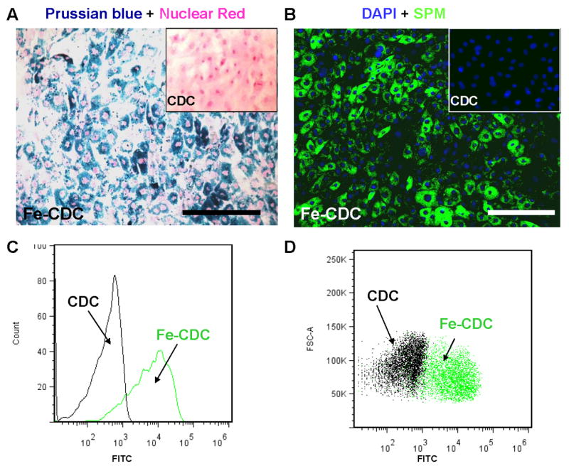
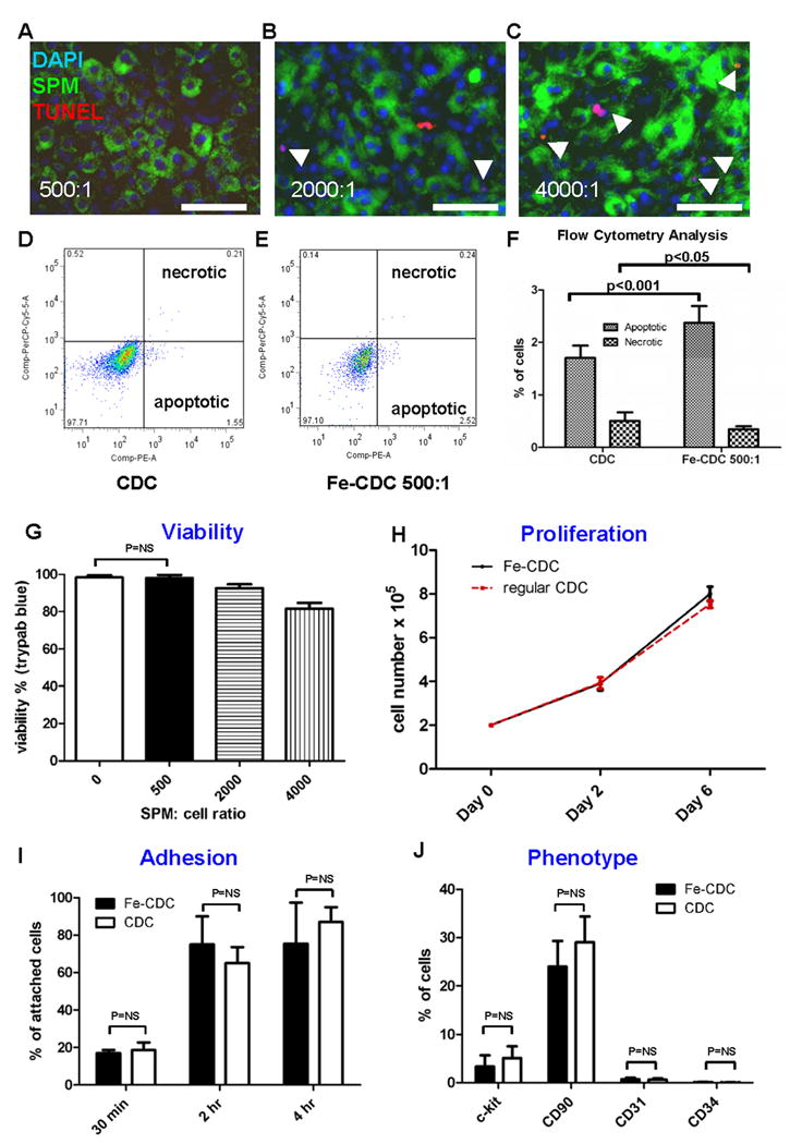
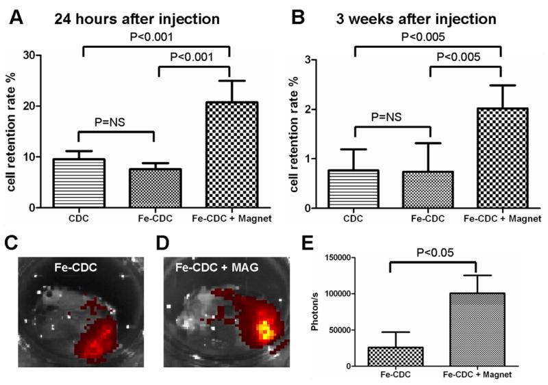
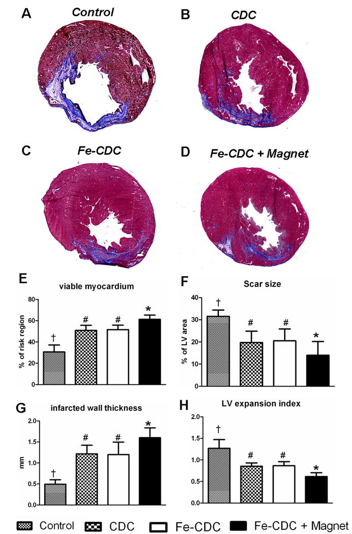
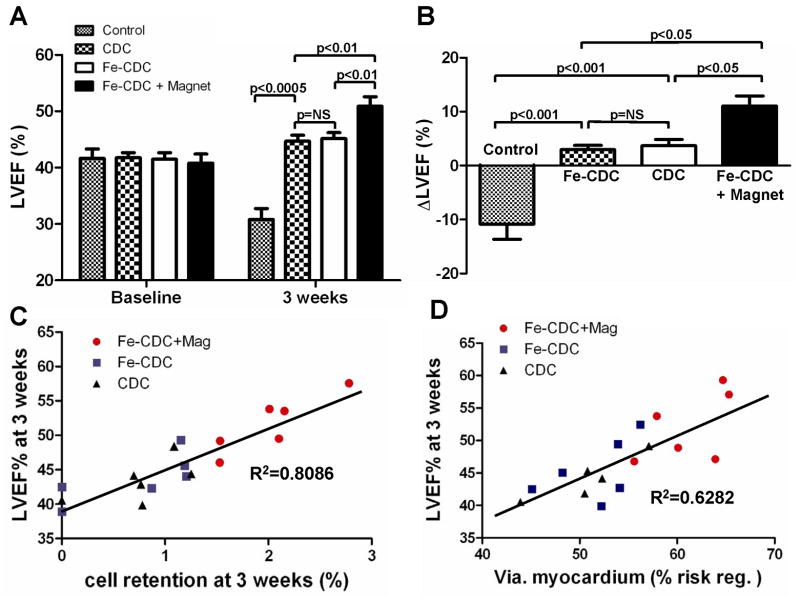
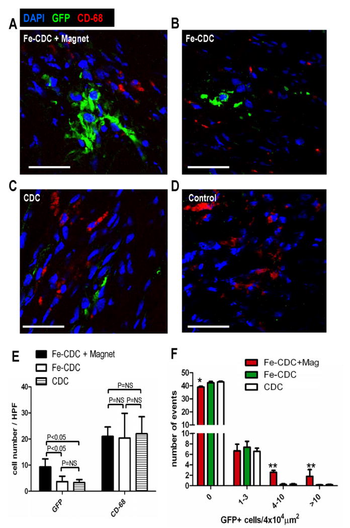
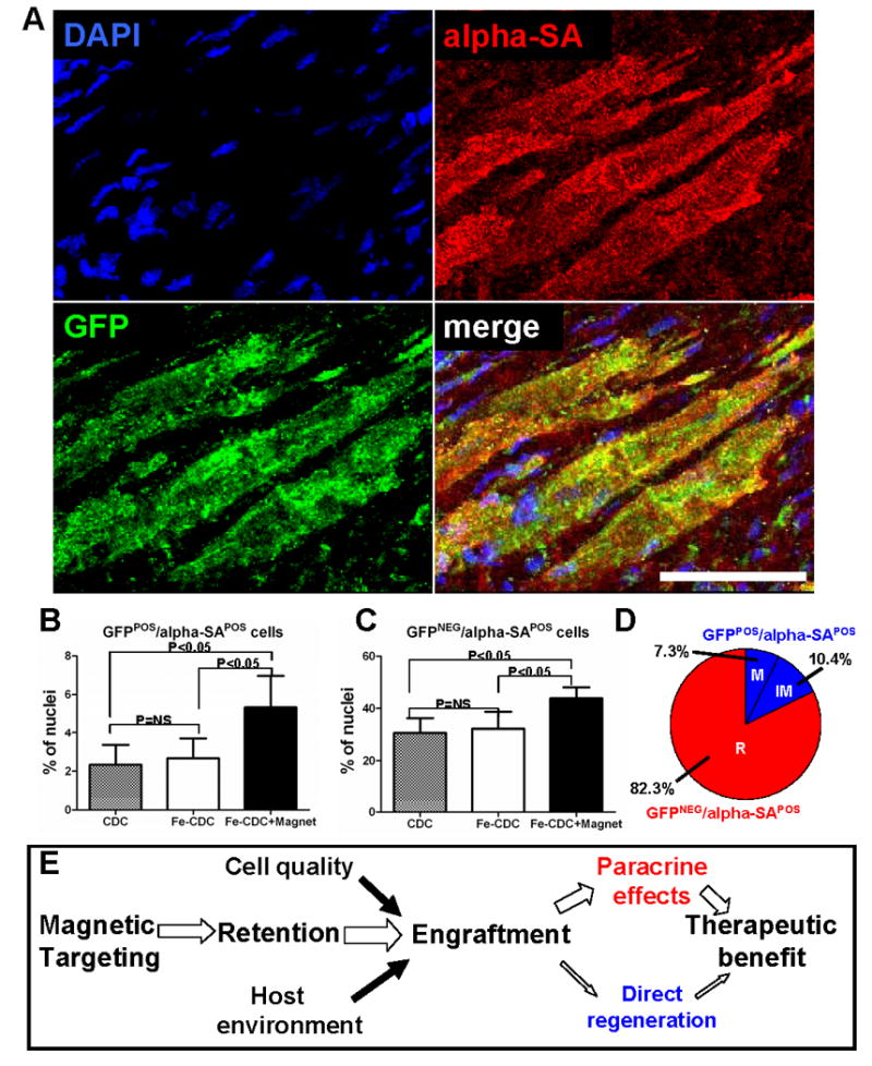
Methods and results: Cardiosphere-derived cells (CDCs) were labeled with superparamagnetic microspheres (SPMs). In vitro studies revealed that cell viability and function were minimally affected by SPM labeling. SPM-labeled rat CDCs were injected intramyocardially, with and without a superimposed magnet. With magnetic targeting, cells were visibly attracted toward the magnet and accumulated around the ischemic zone. In contrast, the majority of nontargeted cells washed out immediately after injection. Fluorescence imaging revealed more retention of transplanted cells in the heart, and less migration into other organs, in the magnetically targeted group. Quantitative PCR confirmed that magnetic targeting enhanced cell retention (at 24 hours) and engraftment (at 3 weeks) in the recipient hearts by approximately 3-fold compared to nontargeted cells. Morphometric analysis revealed maximal attenuation of left ventricular remodeling, and echocardiography showed the greatest functional improvement, in the magnetic targeting group. Histologically, more engrafted cells were evident with magnetic targeting, but there was no incremental inflammation.

Conclusions: Magnetic targeting enhances cell retention, engraftment and functional benefit. This novel method to improve cell therapy outcomes offers the potential for rapid translation into clinical applications.


Conflict of interest statement

Disclosures: Dr. Marbán is a founder and equity holder in Capricor Inc. Capricor provided no funding for the present study. The remaining authors report no conflicts.

Figures

Figure 1
Figure 1
SPM labeling of CDCs. A, rat CDCs were co-incubated with SPMs for 24 hours at a 500:1 SPM: cell ratio. The cells were then fixed, stained for Prussian Blue (iron) and counter-stained with nuclear red. B, CDCs were labeled with dragon-green-conjugated SPMs for 24 hours and then examined by fluorescence microscopy. Non-labeled cells did not express Prussian Blue or Dragon-green fluorescence (Insets, A&B). C and D, representative flow cytometry histogram and dot plots of SPM-labeled (green) and non-labeled CDCs (black). Bars = 100 μm in A and B.
Figure 2
Figure 2
Effects of SPM labeling on cell death and function. A-C, microscopy images of TUNEL staining (Red: apoptotic cells; green: SPMs; blue: nuclei). CDCs were co-incubated with SPMs for 24 hours at varying SPM:cell ratios: 500:1 (A); 2000:1 (B); 4000:1 (C). Apoptotic cells (red color) are highlighted with white arrowheads. Bars = 50 μm. D and E, Typical plots of Annexin/7-AAD flow cytometry from non-labeled CDCs (D) and SPM-labeled CDCs (E). F, quantification of apoptotic and necrotic cells by flow cytometry (n=9 for CDC; n=8 for Fe-CDC). CDCs were labeled with SPMs for 24 hours and then examined for viability and function. G, viability of SPM-labeled CDCs assessed by Trypan Blue exclusion. Viability decrease was only observed in the 2000:1 and 4000:1 dosage groups, but not in the 500:1 group. H, proliferation of Fe-CDCs (labeled at 500:1 SPMs) compared with that of control CDCs (n=4). Cell counts at Day 0, 2 and 6 were equivalent in the two groups. I, adhesion potency of Fe-CDCs (labeled with 500:1 SPMs) compared with that of control CDCs (n=3). Attached cell numbers at 30 min, 2 hours and 4 hours were not statistically different in the two groups. J, phenotypic markers c-kit, CD90, CD31 and CD34 from Fe-CDCs (n=8) were compared to those from control CDCs (n=9). No statistical differences were detected for any of those markers.
Figure 3
Figure 3
Magnetic targeting increases short-term cell retention in the hearts and reduces off-target migration. A and B: Representative images of hearts from the Fe-CDC and Fe-CDC + Magnet group 24 hours after cell injection. Cells are visible as a yellow-brown area in the Fe-CDC + Magnet group (B; red arrow) but not in the Fe-CDC group (A). C-K: Representative fluorescence imaging of organs harvested at 24 hours after cell injection. CDCs were labeled with flash-red-conjugated SPMs. Exposure time was set at the same level for each imaging procedure. More fluorescence was detected in a heart from the Fe-CDC + Magnet group (F) than in the Fe-CDC group (C). Red fluorescence signals were detectable in the lungs and spleens, but less so in the lungs and spleens from the Fe-CDC + Magnet group (G and H) than in those from the Fe-CDC group (D and E). As a negative control, excised organs from the CDC group (animals were injected with non-labeled cells) were also imaged; no signals were detected from any such organs (I-K).
Figure 4
Figure 4
Effects of magnetic targeting on short-term cell retention and long-term cell engraftment. A, female animals (n=6) were sacrificed 24 hours after cell injection. Donor male cells persistent in the female hearts were detected by quantitative PCR for the SRY gene. B, similar PCR experiment performed 3 weeks after injection. C and D, CDCs were labeled with flash-red-conjugated SPMs and then injected into animals with (D) or without (C) magnetic targeting. At 3 weeks after injection, representative hearts from both groups (n=3) were harvested and imaged for detection of flash-red fluorescence. More fluorescence is evident in the Fe-CDC+Magnet heart. E, fluorescence intensities (photon/s) from a fixed region of interest (ROI) measured with the Xenogen software.
Figure 5
Figure 5
Morphometric heart analysis. A-D, Representative Masson's trichrome-stained myocardial sections from a subgroup of animals at 3 weeks after treatment (n=6 for Fe-CDC+Magnet and Fe-CDC; n=5 for CDC and Control). Scar tissue and viable myocardium are identified in blue and red color, respectively. E-H, quantitative analysis and LV morphometric parameters (for definition and calculation methods, please see Supplemental Materials-Detailed Methods). † indicates P<0.05 when compared to any other groups. # indicates P=NS. * indicates P<0.05 when compared to any other groups.
Figure 6
Figure 6
Magnetic targeting enhances functional benefit of CDC transplantation. A, left ventricular ejection fraction (LVEF) measured by echocardiography at baseline and 3 weeks after cell injection (n=12 for Fe-CDC+Magnet and Fe-CDC; n=11 for CDC; n=9 for Control). Baseline LVEFs were indistinguishable among the four groups. 2-tailed paired student t-test revealed that all three cell-treated groups had LVEF improvement, while the LVEF from the control group decreased from baseline. The functional improvement was greater in the Fe-CDC+Magnet group than in the others. B, Changes of LVEF from baseline in each group. Values are expressed as mean ± S.E.M. C & D, 3-week LVEFs were plotted against 3-week cell retention rates and viable myocardium in the risk region, from each animal in each group for which both sets of data were available. Linear regression was performed.
Figure 7
Figure 7
Histological analysis of cell engraftment and inflammatory response. At 3 weeks after cell transplantation, hearts from representative animals (n=5-6) in each group were harvested and frozen-sectioned for histological analysis. Sections from different depths of the heart were stained for CD-68 (macrophages) and counter-stained with DAPI. Confocal imaging was performed for simultaneous detection of transplanted cells (GFP; green) and macrophages (CD-68; red): A, Fe-CDC+magnet; B, Fe-CDC; C, CDC; D, Control. Bars=100 μm. E, GFP-positive cell and macrophage numbers from 6 randomly-selected high power fields (3 from infarct area and 3 from peri-infarct area) on each section were quantified. F, GFP-positive cells in 50 randomly selected fields (4×104μm2) were counted. The number of events was plotted against varying GFP-positive cell numbers. ** and * indicates P<0.01 and P<0.05 respectively, when compared to the CDC or Fe-CDC group.
Figure 8
Figure 8. Cardiac differentiation of transplanted CDCs
A, representative confocal micrographs from a heart in the Fe-CDC + Magnet group showing cells expressing GFP (green) and alpha-SA (red). The colocalization of GFP with alpha-SA indicates that transplanted CDCs participated in regeneration of myocardium, differentiating into a cardiomyocyte phenotype. B and C, quantification of the density of GFPPOS/alpha-SAPOS and GFPNEG/alpha-SAPOS cells in the regions where GFP cells engrafted. D, The percentage distribution of recipient and donor myocytes (both mature and immature) in the increment from the Fe-CDC group to the Fe-CDC+Magnet group is quantified. “M”=mature donor-derived cardiomyocytes; “IM”=immature donor-derived cardiomyocytes; “R”=recipient-derived cardiomyocytes. E, potential mechanism of magnetic targeting-enhanced cell therapy. Bar = 50 μm.

Similar articles

Cited by

References

    1. Wollert KC, Drexler H. Clinical Applications of Stem Cells for the Heart. Circ Res. 2005;96:151–163. - PubMed
    1. Fukushima S, Varela-Carver A, Coppen SR, Yamahara K, Felkin LE, Lee J, Barton PJR, Terracciano CMN, Yacoub MH, Suzuki K. Direct Intramyocardial But Not Intracoronary Injection of Bone Marrow Cells Induces Ventricular Arrhythmias in a Rat Chronic Ischemic Heart Failure Model. Circulation. 2007;115:2254–2261. - PubMed
    1. Robey TE, Saiget MK, Reinecke H, Murry CE. Systems approaches to preventing transplanted cell death in cardiac repair. Journal of Molecular and Cellular Cardiology. 2008;45:567–581. - PMC - PubMed
    1. Ye L, Haider HK, Guo C, Sim EKW. Cell-Based VEGF Delivery Prevents Donor Cell Apoptosis After Transplantation. The Annals of Thoracic Surgery. 2007;83:1233–1234. - PubMed
    1. Zhang M, Methot D, Poppa V, Fujio Y, Walsh K, Murry CE. Cardiomyocyte Grafting for Cardiac Repair: Graft Cell Death and Anti-Death Strategies. Journal of Molecular and Cellular Cardiology. 2001;33:907–921. - PubMed

Publication types