

Case report of Acremonium intraocular infection after cataract extraction
- PMID: 20379462
- PMCID: PMC2850999
- DOI: 10.3341/kjo.2010.24.2.119
Case report of Acremonium intraocular infection after cataract extraction
Abstract
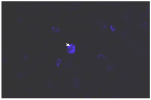
A 64-year-old woman was referred to our clinic for the treatment of chronic uveitis in her left eye, which had started two weeks after an uncomplicated cataract extraction. She was treated with topical steroids with an initially good response, yet she subsequently developed severe inflammation and plaque-like material around the intraocular lens, despite continuous steroid therapy. She underwent pars plana vitrectomy, smear and culture of the aqueous and vitreous fluids, and intravitreal antibiotic injection under the impression of Propionibacterium acne (P. acne) endophthalmitis. As a result of the smear and culture of the vitreous fluid identified as an Acremonium species, she was treated with intravenous amphotericin B injections for five days, followed by oral voriconazole administration. During the post-operative 18-month follow-up, she was stable without significant relapse of uveitis. In this case, the best correction of visual acuity was an improvement from 20/40 to 20/20.
Keywords: Acremonium; Cataract extraction; Endophthalmitis; Propionibacterium.
Conflict of interest statement
No potential conflict of interest relevant to this article was reported.
Figures


Similar articles
-
Management of postoperative Acremonium endophthalmitis.Ophthalmology. 1996 May;103(5):749-56. doi: 10.1016/s0161-6420(96)30620-9. Ophthalmology. 1996. PMID: 8637683
-
Delayed-onset endophthalmitis following cataract surgery caused by Acremonium strictum.Ophthalmic Surg Lasers Imaging. 2005 Nov-Dec;36(6):506-7. Ophthalmic Surg Lasers Imaging. 2005. PMID: 16355956
-
Use of voriconazole in the surgical management of chronic postoperative fungal endophthalmitis.Ophthalmic Surg Lasers Imaging. 2009 Jul-Aug;40(4):425-31. doi: 10.3928/15428877-20096030-16. Ophthalmic Surg Lasers Imaging. 2009. PMID: 19634753
-
A Review of the Role of Intravitreal Corticosteroids as an Adjuvant to Antibiotics in Infectious Endophthalmitis.Ocul Immunol Inflamm. 2018;26(3):461-468. doi: 10.1080/09273948.2016.1245758. Epub 2016 Nov 16. Ocul Immunol Inflamm. 2018. PMID: 27849402 Free PMC article. Review.
-
Fusarium Causing Recalcitrant Post-operative endophthalmitis-Report of a Case with Review of Literature.Ocul Immunol Inflamm. 2022 May 19;30(4):989-991. doi: 10.1080/09273948.2020.1830124. Epub 2021 Oct 12. Ocul Immunol Inflamm. 2022. PMID: 34637660 Review.
Cited by
-
Pseudomonas aeruginosa endophthalmitis masquerading as chronic uveitis.Indian J Ophthalmol. 2013 Jun;61(6):309-10. doi: 10.4103/0301-4738.114122. Indian J Ophthalmol. 2013. PMID: 23803484 Free PMC article.
-
Acremonium species: a review of the etiological agents of emerging hyalohyphomycosis.Mycopathologia. 2010 Dec;170(6):361-75. doi: 10.1007/s11046-010-9334-1. Epub 2010 Jun 25. Mycopathologia. 2010. PMID: 20577905 Review.
-
Post-operative endophthalmitis caused by Acremonium falciforme with orbital and extra-orbital involvement following combined cataract and glaucoma surgery: a case report.J Med Case Rep. 2014 Nov 19;8:373. doi: 10.1186/1752-1947-8-373. J Med Case Rep. 2014. PMID: 25406374 Free PMC article.
References
-
- Meredith TA. Vitrectomy for infectious endophthalmitis. In: Ryan SJ, Hinton DR, Schachat AP, Wilkinson CP, editors. Retina. 4th ed. Vol. 3. Philadelphia: Mosby; 2006. pp. 2260–2261.
-
- Scott IU, Flynn HW, Jr, Miller D. Delayed-onset endophthalmitis following cataract surgery caused by Acremonium strictum. Ophthalmic Surg Lasers Imaging. 2005;36:506–507. - PubMed
-
- Weissgold DJ, Maguire AM, Brucker AJ. Management of postoperative Acremonium endophthalmitis. Ophthalmology. 1996;103:749–756. - PubMed
-
- Cameron JA, Badawi EM, Hoffman PA, Tabara KF. Chronic endophthalmitis caused by Acremonium falciforme. Can J Ophthalmol. 1996;31:367–368. - PubMed
-
- Fridkin SK, Kremer FB, Bland LA, et al. Acremonium kiliense endophthalmitis that occurred after cataract extraction in an ambulatory surgical care and was traced to an environmental reservoir. Clin Infect Dis. 1996;22:222–227. - PubMed
Publication types
MeSH terms
Substances
LinkOut - more resources
Full Text Sources
Research Materials
Miscellaneous
