

Characterization of the anatomic location of the pituitary stalk and its relationship to the dorsum sellae, tuberculum sellae and chiasmatic cistern
- PMID: 20379467
- PMCID: PMC2851087
- DOI: 10.3340/jkns.2010.47.3.169
Characterization of the anatomic location of the pituitary stalk and its relationship to the dorsum sellae, tuberculum sellae and chiasmatic cistern
Abstract
Objective: The normal anatomic relationships characteristic of the pituitary stalk area were previously thought to involve only one location. The purpose of this study was to re-evaluate the anatomic location of the pituitary stalk and possible varying locations in relation to the tuberculum sellae and dorsum sellae using morphometric evaluation and anatomic dissection of human cadaveric specimens. The surgical implications of the variations are discussed.
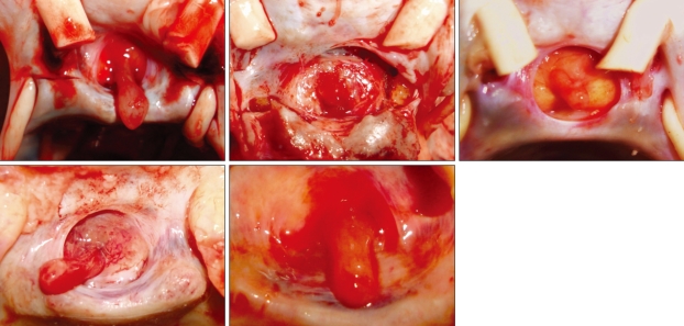
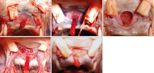
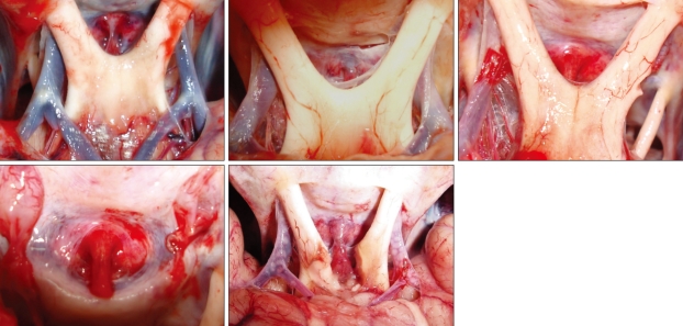
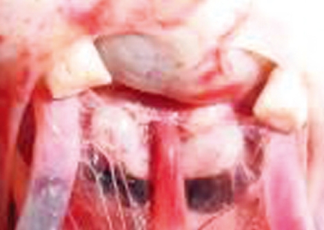
Methods: The calvaria were removed via routine autopsy dissections, and the brains were removed from the skull while preserving the pituitary stalk. The diaphragma sellae, tuberculum sellae, and the location of the pituitary stalk were examined in 60 human cadaveric heads obtained from fresh adult cadavers. Empty sellae were excluded.
Results: The openings of the diaphragma sellae averaged 6.62 +/- 1.606 mm (range, 3-9 mm). The distance between the tuberculum sellae and the posterior part of the pituitary stalk was 1 to 8 mm. The upper face of the diaphragma sellae appeared flat in 26 (43%), concave in 24 (40%), and convex in 6 cases (10%), with a prominent tuberculum sellae in 4 cases (7%). The location of the chiasm was normal in 47 cases (78%), with a prefixed chiasm in 3 cases (5%) and a postfixed chiasm (17%) in the 10 cases. Four cadaver specimens had prominent tuberculum sellae and other parameters were not evaluated.
Conclusion: When opening the chiasmatic cistern, neurosurgeons should be aware about the relationship between the pituitary stalk and the surrounding structures to prevent inadvertent injury to the pituitary stalk.
Keywords: Chiasmatic cistern; Location; Pituitary stalk; Sellae.
Figures
References
-
- Ahmadi H, Larsson EM, Jinkins JR. Normal pituitary gland : coronal MR imaging of infundibular tilt. Radiology. 1990;177:389–392. - PubMed
-
- Amar AP, Weiss MH. Pituitary anatomy and physiology. Neurosurg Clin N Am. 2003;14:11–23. - PubMed
-
- Campero A, Martins C, Yasuda A, Rhoton AL., Jr Microsurgical anatomy of the diaphragma sellae and its role in directing the pattern of growth of pituitary adenomas. Neurosurgery. 2008;62:717–723. discussion 717-723. - PubMed
-
- Carmel PW. Surgical syndromes of the hypothalamus. Clin Neurosurg. 1980;27:133–159. - PubMed
-
- Carmel PW, Antunes JL, Ferin M. Collection of blood from the pituitary stalk and portal veins in monkeys, and from the pituitary sinusoidal system of monkey and man. J Neurosurg. 1979;50:75–80. - PubMed
LinkOut - more resources
Full Text Sources
