

Review
doi: 10.1007/s12350-010-9223-5.
Assessment of myocardial perfusion and function with PET and PET/CT
Affiliations
- PMID: 20379862
- PMCID: PMC2871404
- DOI: 10.1007/s12350-010-9223-5
Item in Clipboard
Review
Assessment of myocardial perfusion and function with PET and PET/CT
J Nucl Cardiol.
2010 Jun.
No abstract available
Figures
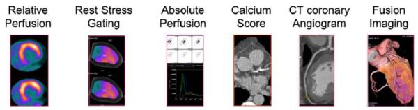
Demonstrates the comprehensive imaging data (relative perfusion, rest and stress gated data, absolute perfusion, calcium score, CT coronary angiogram, and fused PET and CT coronary angiogram images) obtained by a list mode acquisition in a case of combined PET and CT coronary angiography.
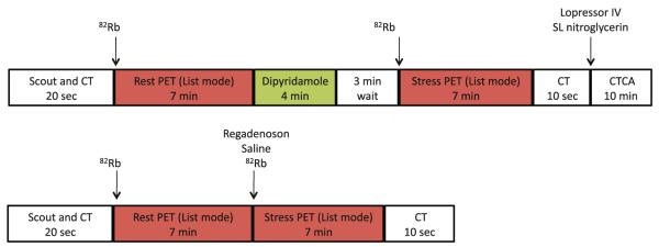
A sample protocol for clinical cardiac PET/CT imaging with 82Rb takes ~25 minutes. The use of Regadenoson stress makes the protocol ultrashort with completion of rest and stress imaging in ~17 minutes. CT, CT scan for attenuation correction; CTCA, CT coronary angiography; 82Rb, 82Rubidium.
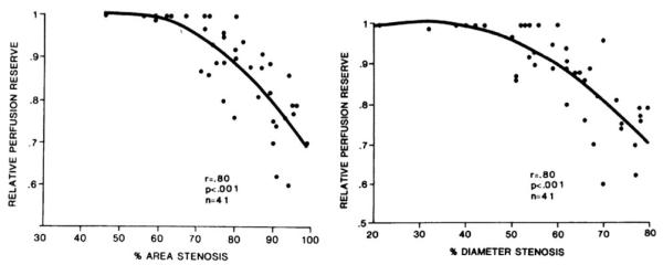
Relative PET perfusion reserve as a tool to assess physiological significance of coronary stenoses. Reproduced with permission from Goldstein et al.
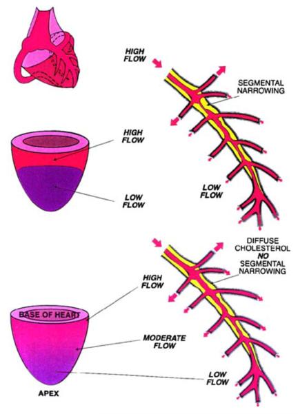
Schematic demonstrating a discrete defect from a segmental coronary stenosis (top), in comparison to the gradual apex to base gradient that may be evident in cases of diffuse CAD. Reproduced with permission from Gould et al.
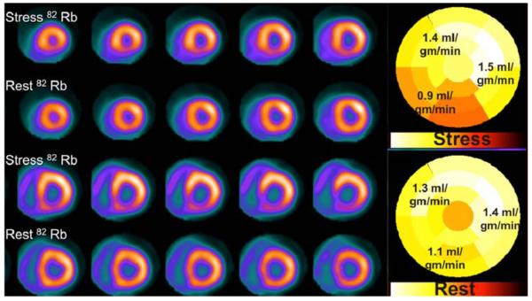
Relative myocardial perfusion images demonstrate inferior and inferoseptal ischemia, while absolute myocardial perfusion is globally reduced, suggesting balanced ischemia.
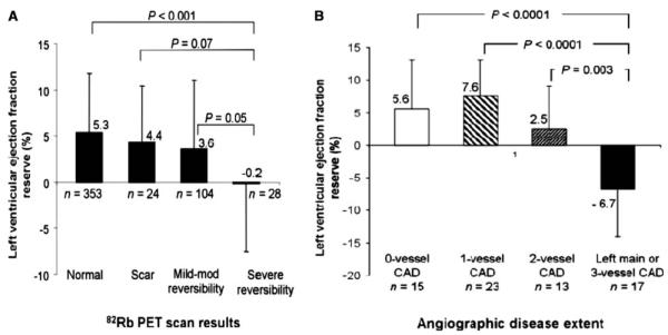
Bar graphs demonstrating the relationship between LVEF reserve (peak stress minus rest LVEF) and the magnitude of stress-induced perfusion abnormalities (A) and the extent of angiographic CAD (>70% stenosis) (B). Reproduced with permission from Dorbala et al.
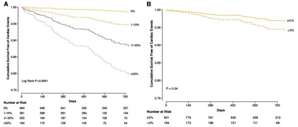
(A) Risk adjusted survival curves demonstrating event free survival based on percent myocardium abnormal. Survival was excellent in patients with normal 82Rb MPI (0% abnormal), and progressively worse survival was noted for patients with mild (1-10% abnormal), moderate (11-20% abnormal), or severely abnormal (≥20% abnormal) scans. (B) Risk adjusted survival curves demonstrating worse event free survival for patients with LVEF reserve <0%. Reproduced with permission from Dorbala et al.
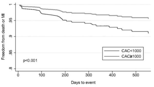
Risk adjusted survival curves in patients with nonischemic relative PET scans demonstrating worse event free survival in patients with a calcium score (CAC) of ≥1000 compared to CAC <1000. Reproduced with permission from Schenker et al.
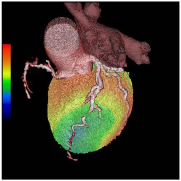
A hybrid PET CTA examination in a patient with significant LAD ischemia. CT coronary angiogram image is overlaid on volume rendered stress 82Rb perfusion image. CT coronary angiogram was performed to evaluate the patency/caliber of the distal LAD (beyond a known chronic total occlusion of the mid LAD) for consideration of coronary artery bypass surgery.
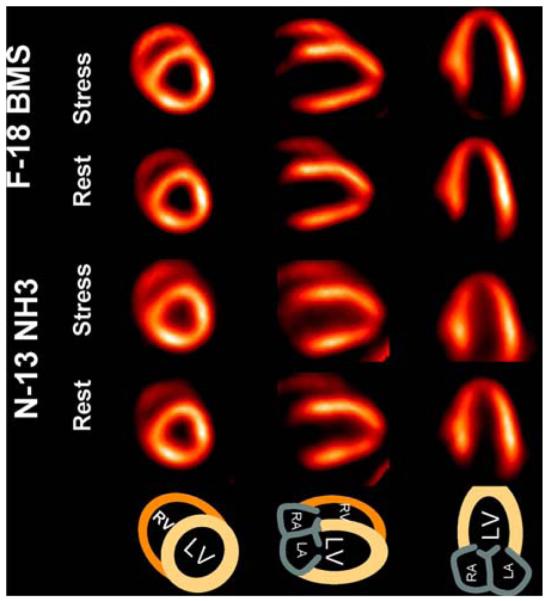
Myocardial perfusion images using F-18 BMS showing comparable or better image quality compared to 13N ammonia images. Reproduced with permission from Nekolla et al.
Similar articles
-
Myocardial perfusion PET/CT to evaluate known and suspected coronary artery disease.Q J Nucl Med Mol Imaging. 2010 Apr;54(2):145-56. Q J Nucl Med Mol Imaging. 2010. PMID: 20588211 Review.
-
Cardiac PET imaging for the detection and monitoring of coronary artery disease and microvascular health.JACC Cardiovasc Imaging. 2010 Jun;3(6):623-40. doi: 10.1016/j.jcmg.2010.04.007. JACC Cardiovasc Imaging. 2010. PMID: 20541718 Review.
-
Computed tomography myocardial perfusion vs 15O-water positron emission tomography and fractional flow reserve.Eur Radiol. 2017 Mar;27(3):1114-1124. doi: 10.1007/s00330-016-4404-5. Epub 2016 Jun 22. Eur Radiol. 2017. PMID: 27334015 Free PMC article.
-
PET measurement of absolute myocardial blood flow and LV function in dilated cardiomyopathy.JACC Cardiovasc Imaging. 2011 May;4(5):557-60. doi: 10.1016/j.jcmg.2010.06.020. JACC Cardiovasc Imaging. 2011. PMID: 21565745 No abstract available.
-
Semi-quantitative myocardial perfusion measured by computed tomography in patients with refractory angina: a head-to-head comparison with quantitative rubidium-82 positron emission tomography as reference.Clin Physiol Funct Imaging. 2017 Sep;37(5):481-488. doi: 10.1111/cpf.12322. Epub 2015 Dec 2. Clin Physiol Funct Imaging. 2017. PMID: 26625937
Cited by
-
PET/CT Myocardial Perfusion Imaging Acquisition and Processing: Ten Tips and Tricks to Help You Succeed.Curr Cardiol Rep. 2021 Mar 11;23(5):39. doi: 10.1007/s11886-021-01476-5. Curr Cardiol Rep. 2021. PMID: 33694057 Review.
-
The role of cardiac PET in diagnosis and prognosis of patients with ischemia with no obstructive coronary arteries (INOCA).Am Heart J Plus. 2024 May 18;43:100399. doi: 10.1016/j.ahjo.2024.100399. eCollection 2024 Jul. Am Heart J Plus. 2024. PMID: 38828445 Free PMC article.
-
Advances in Multimodality Cardiovascular Imaging in the Diagnosis of Heart Failure With Preserved Ejection Fraction.Front Cardiovasc Med. 2022 Mar 9;9:758975. doi: 10.3389/fcvm.2022.758975. eCollection 2022. Front Cardiovasc Med. 2022. PMID: 35355965 Free PMC article. Review.
-
Value of PET ECG gating in a cross-validation study of cardiac function assessment by PET/MR imaging.J Nucl Cardiol. 2023 Jun;30(3):1050-1060. doi: 10.1007/s12350-022-03105-2. Epub 2022 Sep 30. J Nucl Cardiol. 2023. PMID: 36180767 Free PMC article.
-
Current Concepts and Future Applications of Non-Invasive Functional and Anatomical Evaluation of Coronary Artery Disease.Life (Basel). 2022 Nov 7;12(11):1803. doi: 10.3390/life12111803. Life (Basel). 2022. PMID: 36362957 Free PMC article. Review.
References
-
- Beller GA, Bergmann SR. Myocardial perfusion imaging agents: SPECT and PET. J Nucl Cardiol. 2004;11:71–86. - PubMed
-
- Rimoldi OE, Camici PG. Positron emission tomography for quantitation of myocardial perfusion. J Nucl Cardiol. 2004;11:482–90. - PubMed
-
- Di Carli MF. Advances in positron emission tomography. J Nucl Cardiol. 2004;11:719–32. - PubMed
-
- Bengel FM, Higuchi T, Javadi MS, Lautamaki R. Cardiac positron emission tomography. J Am Coll Cardiol. 2009;54:1–15. - PubMed
-
- Di Carli MF, Dorbala S, Meserve J, El Fakhri G, Sitek A, Moore SC. Clinical myocardial perfusion PET/CT. J Nucl Med. 2007;48:783–93. - PubMed
