. 2010 Apr 9:6:19.
doi: 10.1186/1744-8069-6-19.

Spinal microglial motility is independent of neuronal activity and plasticity in adult mice

Affiliations

Spinal microglial motility is independent of neuronal activity and plasticity in adult mice

Tao Chen et al. Mol Pain. .

Abstract

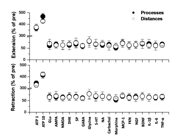
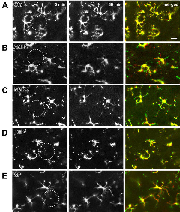
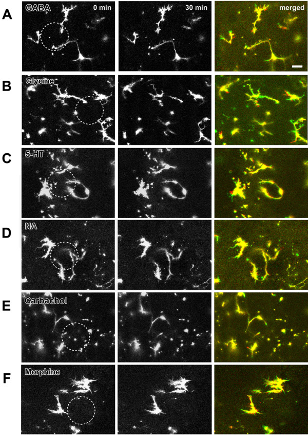
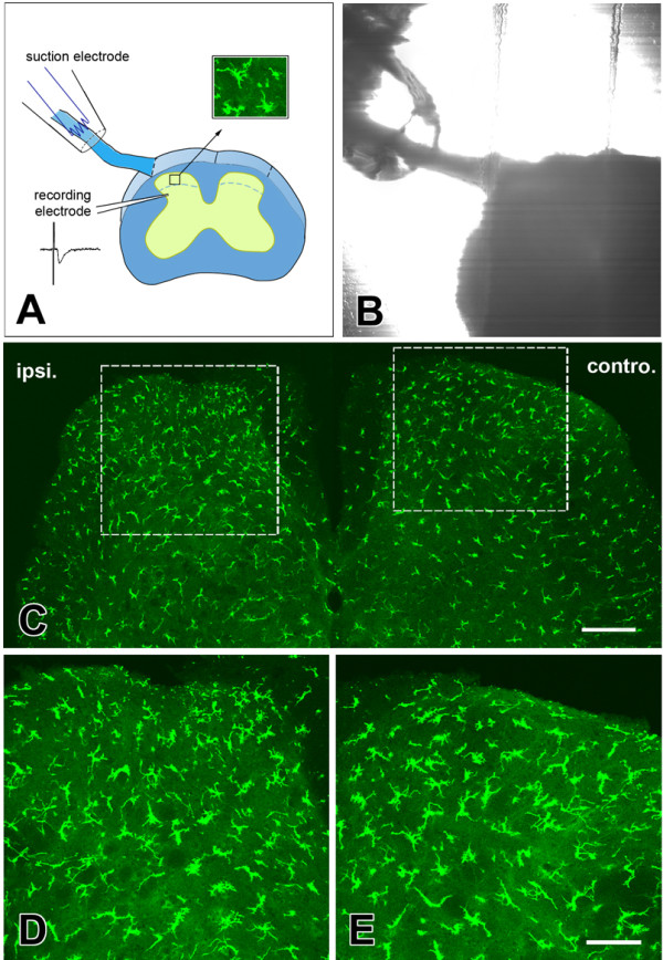
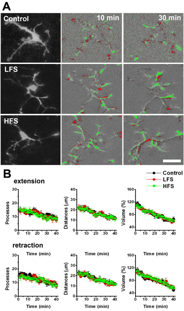
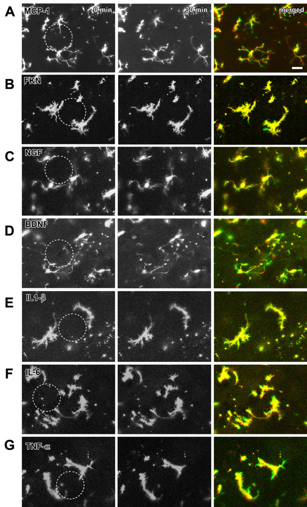
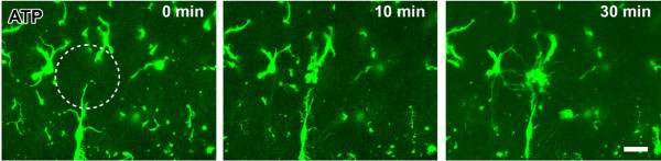
Microglia are the resident macrophages in the central nervous system. In the spinal cord dorsal horn, microglia stay in resting condition during physiological sensory processing, and are activated under pathological conditions such as peripheral nerve injury. In cases such as this, the nearby resting microglia increase their motility and accumulate at the site of injury. However, direct evidence to support that nerve activity can enhance the motility of microglia has not yet to be reported. In this study we investigated whether the activation of spinal microglia under in vivo nerve injury may be mimicked by neuronal activity in the spinal cord slice preparation. We found that local application of spinal excitatory neurotransmitters, such as glutamate and substance P did not cause any change in the motility of microglial cells in the spinal cord dorsal horn. The motility of microglial cells is unlikely modulated by other transmitters, neuromodulators and chemokines, because similar applications such as GABA, serotonin, noradrenaline, carbachol, fractalkine or interleukin did not produce any obvious effect. Furthermore, low or high frequency stimulation of spinal dorsal root fibers at noxious intensities failed to cause any enhanced extension or retraction of the microglia processes. By contrast, focal application of ATP triggered rapid and robust activation of microglial cells in the spinal dorsal horn. Our results provide the first evidence that the activation of microglia in the spinal cord after nerve injury is unlikely due solely to neuronal activity, non-neuronal factors are likely responsible for the activation of nerve injury-related microglial cells in the spinal dorsal horn.


Figures

Figure 1
Figure 1
Summarized effect of different kinds of neurotransmitters, neuromodulators and chemokines on the extension and retraction percentage of microglia in spinal dorsal horn. The ratio of extended or retracted processes and distances of microglia at time 30 min to 0 min after drug application are shown. ATP are applied at 1 or 10 mM; Glu, AMPA, NMDA, DHK, SP, GABA, Glycine, 5-HT, NA, Carbachol and Morphine are applied at 1 mM; MCP-1, FKN, NGF, BDNF, IL-1β, IL-6 and TNF-α are applied at 10 ng/ml. The results of application of 10 mM Glu, AMPA, NMDA, DHK, SP, GABA, Glycine, 5-HT, NA, Carbachol and Morphine and 100 ng/ml MCP-1, FKN, NGF, BDNF, IL-1β, IL-6 and TNF-α are similar. ATP 1, ATP 1 mM; ATP 10, ATP 10 mM; Glu, Glutamate; DHK, dihydrokainate; NA, noradrenalin; SP, Substance P; MCP-1, monocyte chemoattractant protein-1; FKN, fractalkine; NGF, nerve growth factor; BDNF, brain-derived neurotrophic factor; IL-1β, interleukin-1β; IL-6, interleukin-6; TNF-α, tumor necrosis factor-α.
Figure 2
Figure 2
Effect of excitatory neurotransmitters on the motility of microglia in spinal dorsal horn. (A-E). Local application of 1 mM glutamate (Glu), AMPA, NMDA, glutamate uptake inhibitor dihydrokainate (DHK) or substance P (SP) cannot increase the motility of microglia. Dotted circle indicates the center of the drug application area. Microglial motility was imaged every 2 min for 30 min. The first image (0 min, left) and last image (30 min, middle) were shown here. To observe clearer motility, the 2 images were merged (right) and set as green and red for 0 and 30 min, respectively. Therefore the green reflects retracted portions, and the red reflects extended portions, whereas the yellow reflects unaltered portions. Bars equal to 20 μm.
Figure 3
Figure 3
Effect of inhibitory neurotransmitters and neuromodulators on the motility of microglia in spinal dorsal horn. (A-F). Local application of 1 mM GABA, Glycine, 5-HT, noradrenalin (NA), acetylcholine agonist carbachol and morphine had no effect on microglia motility. The merged picture is the overlay of imaging at 0 min (green) and 30 min (red) after drug local application. Note that (F) showed the result recorded on typical amoeboid cells. Bars equal to 20 μm.
Figure 4
Figure 4
Histological results of dorsal root stimuli on the motility of microglia in spinal dorsal horn. (A). Schematic representation of the confocal imaging and field potential recording accompany with dorsal root stimuli; (B). Digitized photomicrographs of the dorsal root stimuli model in a transverse spinal cord section. (C). Observation of microglia in one fixed section of spinal cord after low frequency dorsal root stimuli. Rectangled areas in (C) were magnified in (D) and (E) respectively. Ipsi., ipsilateral part of dorsal root; Contro., controlateral part of dorsal root. Bars equal to 100 μm in (C) and 50 μm in (D) and (E).
Figure 5
Figure 5
Effect of low or high frequency stimuli to the dorsal root on the motility of microglia in spinal dorsal horn. (A). Sample images showing the extension and retraction of microglia in control (top), low frequency stimulation (LFS) (middle) and high frequency stimulation (HFS) induction groups (bottom). Right image is showing the microglia at 10 min and 30 min after imaging. LTP induction was delivered at time 10 min. (B). Extension (top) and retraction (bottom) of microglia in LTP induction groups is similar to that in control group. The number, distance, and percentage of volume of extension or retraction processes were measured. Note that the "rundown" of microglial extension was found in the three groups.
Figure 6
Figure 6
Effect of chemokines on the motility of microglia in spinal dorsal horn. (A-G). Local application of 10 ng/ml monocyte chemoattractant protein-1 (MCP-1), fractalkine (FKN), nerve growth factor (NGF), brain-derived neurotrophic factor (BDNF), interleukin 1β (IL-1β), interleukin 6 (IL-6) or tumor necrosis factor α (TNF-α) had no effect on microglia motility. The merged picture is the overlay of imaging at 0 min (green) and 30 min (red) after drug application. Figs. (E-G) showed the results recorded on amoeboid cells. Bars equal to 20 μm.
Figure 7
Figure 7
Effect of ATP on the motility of microglia in spinal dorsal horn. Local application of ATP (1 mM) induced rapid movement of microglial processes toward the tip of puff pipette in acute spinal cord slices. Three time points of 0 min, 10 and 30 min after ATP application were shown here. Bar equals to 20 μm.

References

    1. Hickey WF. Basic principles of immunological surveillance of the normal central nervous system. Glia. 2001;36:118–124. doi: 10.1002/glia.1101. - DOI - PubMed
    1. Streit WJ, Mrak RE, Griffin WS. Microglia and neuroinflammation: a pathological perspective. J Neuroinflammation. 2004;1:14. doi: 10.1186/1742-2094-1-14. - DOI - PMC - PubMed
    1. Nimmerjahn A, Kirchhoff F, Helmchen F. Resting microglial cells are highly dynamic surveillants of brain parenchyma in vivo. Science. 2005;308:1314–1318. doi: 10.1126/science.1110647. - DOI - PubMed
    1. Inoue K, Tsuda M. Microglia and neuropathic pain. Glia. 2009;57:1469–1479. doi: 10.1002/glia.20871. - DOI - PubMed
    1. Tsuda M, Inoue K, Salter MW. Neuropathic pain and spinal microglia: a big problem from molecules in "small" glia. Trends Neurosci. 2005;28:101–107. doi: 10.1016/j.tins.2004.12.002. - DOI - PubMed

Publication types

MeSH terms