

Results of a multi-institutional benchmark test for cranial CT/MR image registration
- PMID: 20381270
- PMCID: PMC2906611
- DOI: 10.1016/j.ijrobp.2009.10.017
Results of a multi-institutional benchmark test for cranial CT/MR image registration
Abstract
Purpose: Variability in computed tomography/magnetic resonance imaging (CT/MR) cranial image registration was assessed using a benchmark case developed by the Quality Assurance Review Center to credential institutions for participation in Children's Oncology Group Protocol ACNS0221 for treatment of pediatric low-grade glioma.
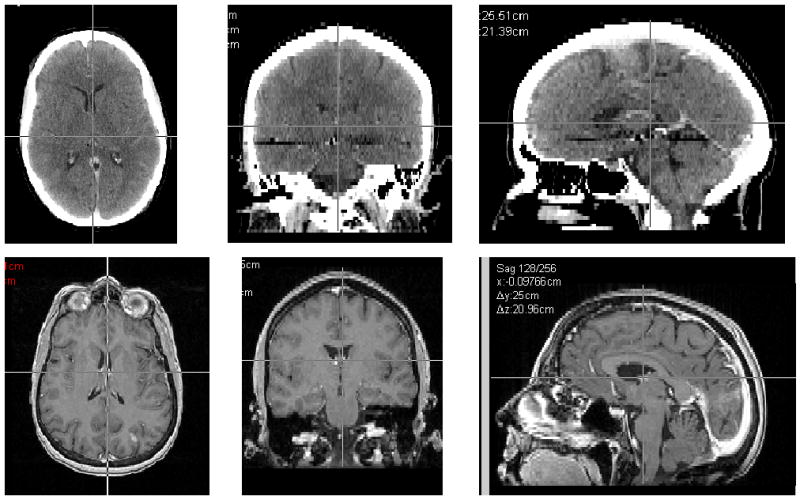
Methods and materials: Two DICOM image sets, an MR and a CT of the same patient, were provided to each institution. A small target in the posterior occipital lobe was readily visible on two slices of the MR scan and not visible on the CT scan. Each institution registered the two scans using whatever software system and method it ordinarily uses for such a case. The target volume was then contoured on the two MR slices, and the coordinates of the center of the corresponding target in the CT coordinate system were reported. The average of all submissions was used to determine the true center of the target.
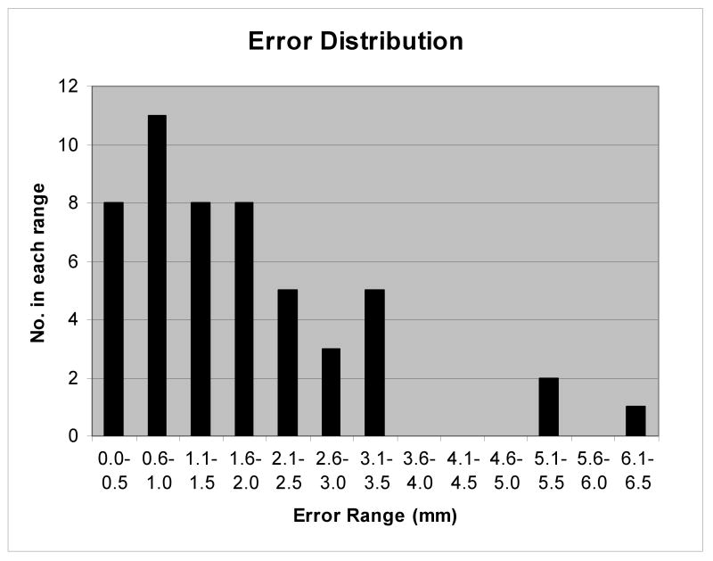
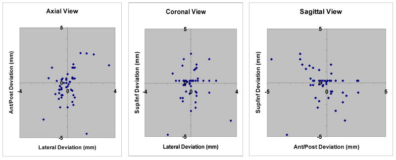
Results: Results are reported from 51 submissions representing 45 institutions and 11 software systems. The average error in the position of the center of the target was 1.8 mm (1 standard deviation = 2.2 mm). The least variation in position was in the lateral direction. Manual registration gave significantly better results than did automatic registration (p = 0.02).
Conclusion: When MR and CT scans of the head are registered with currently available software, there is inherent uncertainty of approximately 2 mm (1 standard deviation), which should be considered when defining planning target volumes and PRVs for organs at risk on registered image sets.
Copyright 2010 Elsevier Inc. All rights reserved.
Conflict of interest statement
Conflicts of Interest Notification: No conflicts of interest exist.
Figures


References
-
- Hamm KD, Surber G, Schmucking M, et al. Stereotactic radiation treatment planning and follow-up studies involving fused multimodality imaging. J Neurosurg. 2004;101(Suppl 3):326–333. - PubMed
-
- Balter JM, Kessler ML. Imaging and alignment for image-guided radiation therapy. J Clin Oncol. 2007;25:931–937. - PubMed
-
- Jin JY, Ryu S, Faber K, et al. 2D/3D image fusion for accurate target localization and evaluation of a mask based stereotactic system in fractionated stereotactic radiotherapy of cranial lesions. Med Phys. 2006;33:4557–4566. - PubMed
-
- Sarkar A, Santiago RJ, Smith R, et al. Comparison of manual vs. automated multimodality (CT-MRI) image registration for brain tumors. Med Dosim. 2005;30:20–24. - PubMed
