. 2010 Jun 1;99(3):117-25.
doi: 10.1016/j.jphotobiol.2010.03.005. Epub 2010 Mar 21.

Inhibition of electron transport chain assembly and function promotes photodynamic killing of Candida

Affiliations

Inhibition of electron transport chain assembly and function promotes photodynamic killing of Candida

Yeissa Chabrier-Roselló et al. J Photochem Photobiol B. .

Abstract

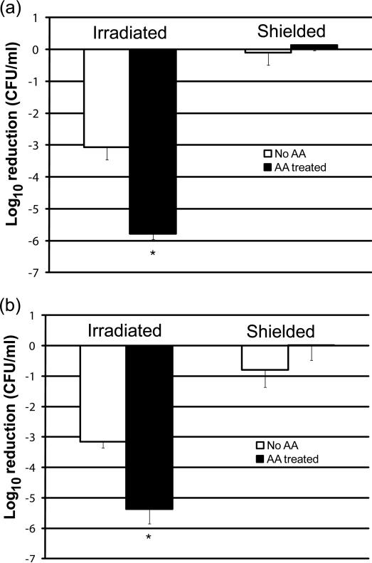
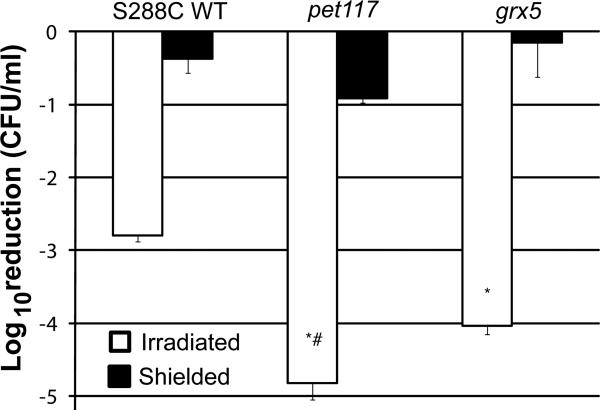
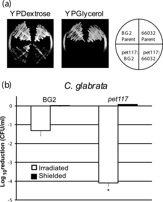
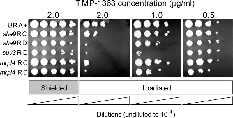
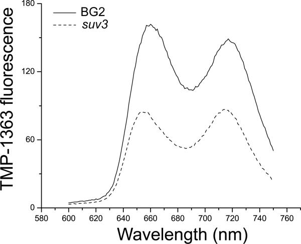
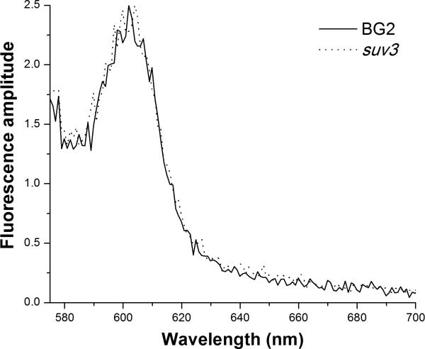
Respiratory deficiency increases the sensitivity of the pathogenic fungi Candida albicans and Candida glabrata to oxidative stress induced by photodynamic therapy (PDT) sensitized by the cationic porphyrin meso-tetra (N-methyl-4-pyridyl) porphine tetra tosylate (TMP-1363). Since disruption of electron transport chain (ETC) function increases intracellular levels of reactive oxygen species in yeast, we determined whether interference with ETC assembly or function increased sensitivity to TMP-1363-PDT in C. albicans, C. glabrata and the non-pathogenic yeast Saccharomyces cerevisiae. Metabolic inhibitor antimycin A and defined genetic mutants were used to identify ETC components that contribute to the sensitivity to PDT. Inhibition of cytochrome bc(1) (Complex III) with antimycin A increases mitochondrial levels of reactive oxygen species. PDT performed following pre-treatment with antimycin A reduced colony forming units (CFU) of C. albicans and C. glabrata by approximately two orders of magnitude relative to PDT alone. A S. cerevisiae mitochondrial glutaredoxin grx5 mutant, defective in assembly of Fe-S clusters critical for Complex III function, displayed increased sensitivity to PDT. Furthermore, C. glabrata and S.cerevisiae mutants in cytochrome c oxidase (Complex IV) synthesis and assembly were also significantly more sensitive to PDT. These included suv3, encoding an ATP-dependent RNA helicase critical for maturation of cytochrome c oxidase subunit transcripts, and pet117, encoding an essential cytochrome c oxidase assembly factor. Following PDT, the reduction in CFU of these mutants was one to two orders of magnitude greater than in their respective parental strains. The data demonstrate that selective inhibition of ETC Complexes III and IV significantly increases the sensitivity of C. albicans, C. glabrata and S. cerevisiae to PDT sensitized with TMP-1363.


Figures

Figure 1
Figure 1. Spectrum of visible radiation from the light box used for PDT
The inset is an expanded view of the region of the spectrum in the wavelength range of 575 - 700 nm that accounts for ~ 67% of the total power emitted from the light source.
Figure 2
Figure 2. Metabolic inhibition of respiratory Complex III by antimycin A increases the sensitivity of C. albicans and C. glabrata to PDT
C. albicans (panel a) and C. glabrata (panel b) were grown overnight to early stationary phase in YPDextrose at 37°C with vigorous aeration, in either the presence (closed bars) or absence (open bars) of the inhibitor of respiratory complex III inhibitor antimycin A (10 μM AA). Organisms were adjusted to 107 cells/mL, incubated with 10 μg/mL of TMP-1363 for 10 min and irradiated at 2.4 J/cm2 with broadband visible light. Untreated organisms and organisms treated with TMP-1363 but shielded from light were used as controls. Organism killing was determined by the colony forming unit (CFU) assay and represented as a log10 reduction compared to untreated organisms. Data represent the mean ± S.D. of three experiments performed in duplicate. Pre-treatment with AA significantly (*) enhanced (p<0.001) the sensitivity of both C. albicans and C. glabrata to PDT compared to untreated organisms.
Figure 3
Figure 3. Genetic evidence that Pet117 and Grx5 participate in sensitivity to PDT in S
Early stationary phase yeast of S. cerevisiae S288C, pet117 and grx5 were incubated with 10 μg/ml TMP-1363 for 10 min and irradiated with a fluence of 2.4 J/cm2 broadband visible light (open bars). Untreated organisms and organisms treated with TMP-1363 but shielded from light (closed bars) were used as controls. Organism killing was determined by the CFU assay and represented as a log10 reduction compared to untreated organisms. Data represent the mean ± S.D. of three experiments performed in triplicate. Both S. cerevisiae respiratory-deficient mutants pet117 and grx5 displayed significantly (*) increased sensitivity to oxidative stress mediated by PDT (p<0.001) compared to parental strain S288C. The pet117 mutant was also significantly (#) more sensitive to PDT (p=0.003) compared to grx5.
Figure 4
Figure 4. C. glabrata pet117 insertion mutants are respiratory deficient and display increased sensitivity to PDT
(a) Representative clones in both the C. glabrata BG2 and C. glabrata 66032 background incubated at 30oC for 48 h are shown. The diagram on the rightshows the organization of the isolates on the YPDextrose and YPGlycerol agar plates. Both transformants show the “petite” phenotype of smaller colony size and reduced growth on YPDextrose, and the putative pet117 insertion mutants are unable to grow on YPGlycerol, confirming their respiratory deficiency (lower quadrants). The parental strains BG2 and 66032 grew well on both media (upper quadrants). (b) Early stationary phase yeast of C. glabrata BG2 and pet117 were treated with 0.5 μg/ml TMP-1363 for 10 min and irradiated at a fluence of 2.4 J/cm2 with broadband visible light (open bars). Untreated organisms and organisms treated with TMP-1363 but shielded from light (closed bars) were used as controls. Sensitivity to PDT was assessed by the CFU assay. Data are expressed as a log10 reduction in CFU compared to untreated organisms, and represent the mean + S.D. of three experiments performed in triplicate. C. glabrata pet117 exhibited significantly (*) enhanced sensitivity to PDT compared to parental strain BG2 (p<0.001).
Figure 5
Figure 5. Selected pathways leading to respiratory deficiency in C. glabrata contribute to increased susceptibility to PDT
To better reveal differences in sensitivity to PDT among the strains, organisms were adjusted to 107 cells/ml, incubated with a range of concentrations (0.5 – 2.0 μg/ml) of TMP-1363 for 10 min and irradiated with a fluence of 2.4 J/cm2 broadband visible light (Irradiated). Organisms treated with TMP-1363 but shielded from light were used as controls (Shielded). Organism killing was determined by spotting 2 μl of ten-fold serial dilutions of treated cell suspensions on YPDextrose plates, followed by incubation at 37°C for 24 h. Representative results are shown. Abbreviations: RC (respiratory-competent); RD (respiratory-deficient).
Figure 6
Figure 6. Comparison of cell-associated photosensitizer levels in C. glabrata parental and respiratory-deficient strains
TMP-1363 steady state fluorescence from wild type C. glabrata BG2 (solid line) and respiratory-deficient mutant suv3 (dashed line) strains. The excitation wavelength was 425 nm. The shapes of the TMP-1363 excitation spectra (not shown) from these two strains were indistinguishable, thus the differences in amplitude reflect significant differences in sensitizer concentration. In this case, the level of TMP-1363 associated with respiratory-deficient mutant C. glabrata suv3 was less than its parent BG2.
Figure 7
Figure 7. Comparison of endogenous fluorescence levels in C. glabrata parental and respiratory-deficient mutant strains
Endogenous fluorescence from wild type C. glabrata BG2 (solid line) and respiratory-deficient mutant suv3 (dashed line) was excited at 500 nm. The spectral shape and amplitude of the emission from these two strains were indistinguishable. The displayed amplitudes were scaled to illustrate the 50-100-fold lower emission intensities of the endogenous fluorescence relative to that of TMP-1363 (Fig. 6).

References

    1. Calderone RA. Candida and Candidiasis. ASM Press; Washington, D.C.: 2002.
    1. Fidel PL, Jr., Vazquez JA, Sobel JD. Candida glabrata: review of epidemiology, pathogenesis, and clinical disease with comparison to C. albicans. Clin. Microbiol. Rev. 1999;12:80–96. - PMC - PubMed
    1. Pfaller MA, Messer SA, Hollis RJ, Jones RN, Doern GV, Brandt ME, Hajjeh RA. Trends in species distribution and susceptibility to fluconazole among blood stream isolates of Candida species in the United States. Diag. Microbiol. Infect. Dis. 1999;33:217–222. - PubMed
    1. Johnson EM, Warnock DW, Luker J, Porter SR, Scully C. Emergence of azole drug resistance in Candida species from HIV-infected patients receiving prolonged fluconazole therapy for oral candidosis. J. Antimicrob. Chemother. 1995;35:103–114. - PubMed
    1. Li L, Redding S, Dongari-Bagtzoglou A. Candida glabrata: an emerging oral opportunistic pathogen. J. Dent. Res. 2007;86:204–215. - PubMed

Publication types

MeSH terms

LinkOut - more resources