

Vasopressin cell groups exhibit strongly divergent responses to copulation and male-male interactions in mice
- PMID: 20382147
- PMCID: PMC4195792
- DOI: 10.1016/j.yhbeh.2010.03.021
Vasopressin cell groups exhibit strongly divergent responses to copulation and male-male interactions in mice
Abstract
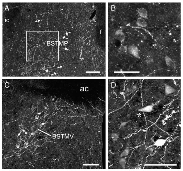
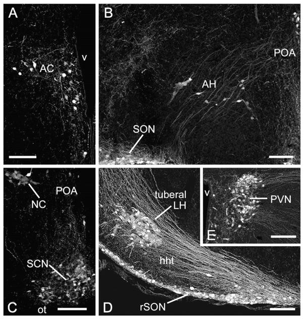
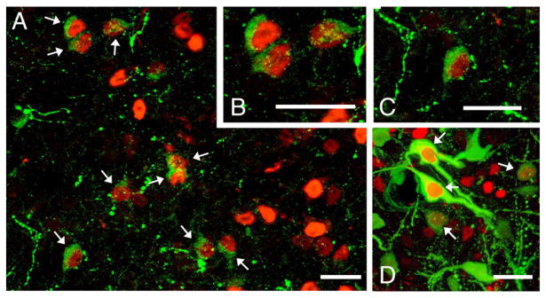
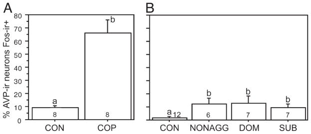
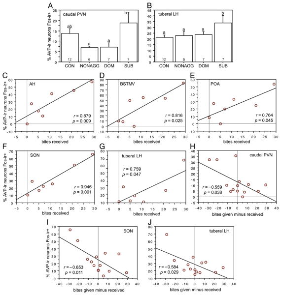
Arginine vasopressin (AVP) and its nonmammalian homolog arginine vasotocin influence social behaviors ranging from affiliation to resident-intruder aggression. Although numerous sites of action have been established for these behavioral effects, the involvement of specific AVP cell groups in the brain is poorly understood, and socially elicited Fos responses have not been quantified for many of the AVP cell groups found in rodents. Surprisingly, this includes the AVP population in the posterior part of the medial bed nucleus of the stria terminalis (BSTMP), which has been extensively implicated, albeit indirectly, in various aspects of affiliation and other social behaviors. We examined the Fos responses of eight hypothalamic and three extra-hypothalamic AVP-immunoreactive (-ir) cell groups to copulation, nonaggressive male-male interaction, and aggressive male-male interaction in both dominant and subordinate C57BL/6J mice. The BSTMP cells exhibited a response profile that was unlike all other cell groups: from a control baseline of approximately 5% of AVP-ir neurons colocalizing with Fos, colocalization increased significantly to approximately 12% following nonaggressive male-male interaction, and to approximately 70% following copulation. Aggressive interactions did not increase colocalization beyond the level observed in nonaggressive male mice. These results suggest that BSTMP neurons in mice may increase AVP-Fos colocalization selectively in response to affiliation-related stimuli, similar to findings in finches. In contrast, virtually all other cell groups were responsive to negative aspects of interaction, either through elevated AVP-Fos colocalization in subordinate animals, positive correlations of AVP-Fos colocalization with bites received, and/or negative correlations of AVP-Fos colocalization with dominance. These findings greatly expand what is known of the contributions of specific brain AVP cell groups to social behavior.
Published by Elsevier Inc.
Figures
Similar articles
-
Hypothalamic vasopressin systems are more sensitive to the long term effects of social defeat in males versus females.Psychoneuroendocrinology. 2015 Jan;51:122-34. doi: 10.1016/j.psyneuen.2014.09.009. Epub 2014 Sep 19. Psychoneuroendocrinology. 2015. PMID: 25306217 Free PMC article.
-
Distinct correlations of vasopressin release within the lateral septum and the bed nucleus of the stria terminalis with the display of intermale aggression.Horm Behav. 2010 Jul;58(2):273-81. doi: 10.1016/j.yhbeh.2010.03.006. Epub 2010 Mar 15. Horm Behav. 2010. PMID: 20298693
-
Differences in intermale aggression are accompanied by opposite vasopressin release patterns within the septum in rats bred for low and high anxiety.Eur J Neurosci. 2007 Dec;26(12):3597-605. doi: 10.1111/j.1460-9568.2007.05974.x. Epub 2007 Dec 4. Eur J Neurosci. 2007. PMID: 18052969
-
Vasopressin and serotonin interactions in the control of agonistic behavior.Psychoneuroendocrinology. 1994;19(5-7):593-601. doi: 10.1016/0306-4530(94)90043-4. Psychoneuroendocrinology. 1994. PMID: 7938357 Review.
-
Nonapeptides and the evolutionary patterning of sociality.Prog Brain Res. 2008;170:3-15. doi: 10.1016/S0079-6123(08)00401-9. Prog Brain Res. 2008. PMID: 18655867 Free PMC article. Review.
Cited by
-
Modulation of social behavior by distinct vasopressin sources.Front Endocrinol (Lausanne). 2023 Feb 13;14:1127792. doi: 10.3389/fendo.2023.1127792. eCollection 2023. Front Endocrinol (Lausanne). 2023. PMID: 36860367 Free PMC article. Review.
-
A vasopressin circuit that modulates mouse social investigation and anxiety-like behavior in a sex-specific manner.Proc Natl Acad Sci U S A. 2024 May 14;121(20):e2319641121. doi: 10.1073/pnas.2319641121. Epub 2024 May 6. Proc Natl Acad Sci U S A. 2024. PMID: 38709918 Free PMC article.
-
Pheromone exposure influences preoptic arginine vasotocin gene expression and inhibits social approach behavior in response to rivals but not potential mates.Brain Behav Evol. 2013;81(3):194-202. doi: 10.1159/000350589. Epub 2013 May 22. Brain Behav Evol. 2013. PMID: 23712040 Free PMC article.
-
Behavioral relevance of species-specific vasotocin anatomy in gregarious finches.Front Neurosci. 2013 Dec 17;7:242. doi: 10.3389/fnins.2013.00242. eCollection 2013. Front Neurosci. 2013. PMID: 24381536 Free PMC article.
-
Mammal-like organization of the avian midbrain central gray and a reappraisal of the intercollicular nucleus.PLoS One. 2011;6(6):e20720. doi: 10.1371/journal.pone.0020720. Epub 2011 Jun 7. PLoS One. 2011. PMID: 21694758 Free PMC article.
References
-
- Beiderbeck DI, Neumann ID, Veenema AH. Differences in intermale aggression are accompanied by opposite vasopressin release patterns within the septum in rats bred for low and high anxiety. Eur J Neurosci. 2007;26:3597–3605. - PubMed
-
- Bielsky IF, Hu SB, Ren X, Terwilliger EF, Young LJ. The V1a vasopressin receptor is necessary and sufficient for normal social recognition: a gene replacement study. Neuron. 2005;47:503–513. - PubMed
-
- Bielsky IF, Hu SB, Szegda KL, Westphal H, Young LJ. Profound impairment in social recognition and reduction in anxiety-like behavior in vasopressin V1a receptor knockout mice. Neuropsychopharmacology. 2004;29:483–493. - PubMed
-
- Bolborea M, Ansel L, Weinert D, Steinlechner S, Pevet P, Klosen P. The bed nucleus of the stria terminalis in the Syrian hamster (Mesocricetus auratus): absence of vasopressin expression in standard and wild-derived hamsters and galanin regulation by seasonal changes in circulating sex steroids. Neuroscience. 2010;165:819–830. - PubMed
Publication types
MeSH terms
Substances
Grants and funding
LinkOut - more resources
Full Text Sources
Miscellaneous
