. 2010 Jun;176(6):2958-71.
doi: 10.2353/ajpath.2010.090838. Epub 2010 Apr 9.

Capturing changes in the brain microenvironment during initial steps of breast cancer brain metastasis

Affiliations

Capturing changes in the brain microenvironment during initial steps of breast cancer brain metastasis

Mihaela Lorger et al. Am J Pathol. 2010 Jun.

Abstract

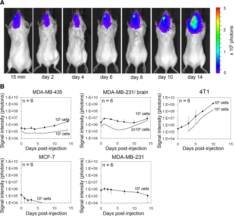
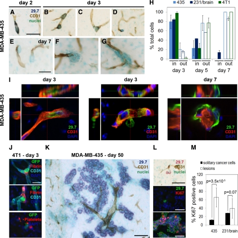
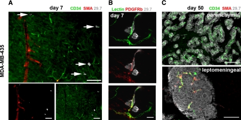
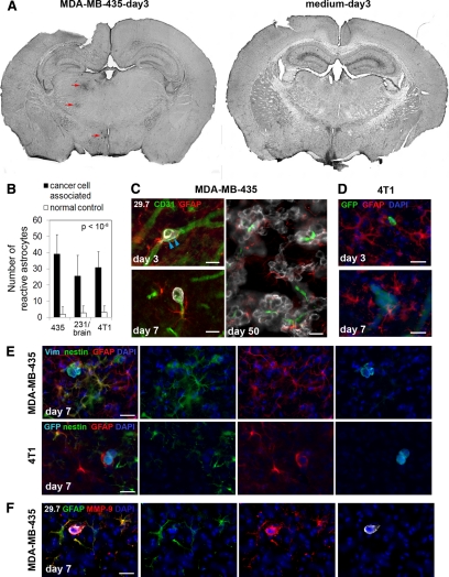
Brain metastases are difficult to treat and mostly develop late during progressive metastatic disease. Patients at risk would benefit from the development of prevention and improved treatments. This requires knowledge of the initial events that lead to brain metastasis. The present study reveals cellular events during the initiation of brain metastasis by breast cancer cells and documents the earliest host responses to incoming cancer cells after carotid artery injection in immunodeficient and immunocompetent mouse models. Our findings capture and characterize heterogeneous astrocytic and microglial reactions to the arrest and extravasation of cancer cells in the brain, showing immediate and drastic changes in the brain microenvironment on arrival of individual cancer cells. We identified reactive astrocytes as the most active host cell population that immediately localizes to individual invading tumor cells and continuously associates with growing metastatic lesions. Up-regulation of matrix metalloproteinase-9 associated with astrocyte activation in the immediate vicinity of extravasating cancer cells might support their progression. Early involvement of different host cell types indicates environmental clues that might codetermine whether a single cancer cell progresses to macrometastasis or remains dormant. Thus, information on the initial interplay between brain homing tumor cells and reactive host cells may help develop strategies for prevention and treatment of symptomatic breast cancer brain metastases.


Figures

Figure 1
Figure 1
Brain colonization by breast cancer cell lines after carotid artery injection in mice monitored by bioluminescence imaging. A: Increase of bioluminescence signal indicates growth of F-luc-tagged MDA-MB-435 cells shown in a representative animal at various time points after injection of 105 cancer cells. B: Survival and growth of F-luc-tagged MDA-MB-435, MDA-MB-231 parental, MDA-MB-231/brain, MCF-7 (injected into immunosuppressed CB17/SCID mice), and 4T1 cells (injected into immunocompetent BALB/c mice) after injection into the left carotid artery followed over time. Only live cells produce signal. Number of injected cells as indicated.
Figure 2
Figure 2
Cancer cell extravasation and growth in the brain. A–G: MDA-MB-435 cells were visualized by anti-human CD44 (mAb 29.7) and blood vessels by CD31 staining using immunohistochemistry. A: An elongated cancer cell within a capillary on day two postinjection. B: Rounding of intravascular cancer cells on day three. C: Cancer cell on day three breaking through the vessel wall during extravasation. D: Extravasated cancer cell on day three. E–G: Extravascular cancer cells on day seven. Scale bars: 50 μm (A–D); 25 μm (E–G). H: Percentage of cancer cells located inside versus outside blood vessels. The quantification was performed for three different cells lines: MDA-MB-435, MDA-MB-231/brain, and 4T1. I: Analysis of early cell location by confocal microscopy. Cancer cells were stained for human CD44 (green) and blood vessels for CD31 (red). An intravascular cell (left panel), a cell in the process of extravasation (middle panel), and extravascular cells (right panel) are shown. J: Association of intravascular 4T1 cancer cells with fibrin and platelets (GPIbα staining) on day three. Scale bar: 25 μm. K: Day 50: Long-term fate of MDA-MB-435 cells was monitored by immunohistochemistry. Intraparenchymal macrometastases grew preferentially around co-opted blood vessels. Scale bar: 200 μm. L: Solitary tumor cells outside blood vessels on day 50 (top panel), detected by anti-human CD44 (mAb 29.7), are mostly negative for Ki-67 (middle panel). In contrast, most cells within lesions as shown in K are Ki-67 positive (bottom panel). Scale bars: 50 μm. M: Quantification of Ki-67-positive cells within the solitary cancer cell population and within the macrometastatic lesions for MDA-MB-435 and MDA-MB-231/brain cells 30 to 50 days postinjection.
Figure 3
Figure 3
Blood vessel types involved in cancer cell extravasation and growth. Blood vessel types were analyzed by immunofluorescence. Representative images for MDA-MB-435 cells are shown. A: Day seven: in the brain parenchyma, cancer cells (white, arrows) arrest and extravasate exclusively from capillaries or postcapillary venules positive for CD34 and lacking smooth muscle cells (no smooth muscle actin (SMA) signal). Scale bars: 100 μm. B: Day seven: blood vessels from which cancer cells (gray) extravasate are surrounded by platelet-derived growth factor receptor β (PDGFRb)-positive pericytes. Top panel: BS-1 lectin; middle panel: anti-PDGFRb; and bottom panel: merge. Scale bar: 20 μm. C: Day 50: intraparenchymal metastases grow around co-opted capillaries lacking smooth muscle cells. Leptomeningeal metastases contain capillaries as well as larger, smooth muscle cell-positive vessels. Scale bars: 100 μm.
Figure 4
Figure 4
Microglial cell responses to invading cancer cells are heterogeneous. Microglial activation in response to cancer cell invasion varies at early as well as late stages, as detected by immunofluorescence analysis. A–G: MDA-MB-435 cells in immunosuppressed mice; diverse microglial responses (green) to incoming cancer cells (red) on day seven (A). Responses include absence of microglial cells (B), presence of hypertrophic stellate activated (D), or amoeboid reactive microglial cells (F). Similarly, on day 50, some macrometastases show no microglial involvement (C) or contain stellate (E) or amoeboid microglia (G). Scale bars: 100 μm (A); 50 μm (B–G). H–K: 4T1 cells in immunocompetent mice; H: distribution of activated microglia (red) in the mouse brain seven days after carotid artery injection of cancer cells (left panel) or medium alone (right panel). White arrowheads mark the GFP-labeled cancer cells (green). Diverse microglial responses to cancer cells include absence of microglial cells (I), presence of hypertrophic stellate (J), or reactive amoeboid microglial cells (K). Scale bar: 50 μm.
Figure 5
Figure 5
Cancer cell invasion induces strong astrocytic responses. Astrocytes were investigated by immunofluorescence staining. A: Left, On day three after cancer cell injection into the left carotid artery, GFAP in astrocytes is already up-regulated strongly in the vicinity of intravascular arrested cancer cells (MDA-MB-435, red arrows). Astrocyte activation can be detected in the left hemisphere in brain overview sections, whereas the corresponding area of the contralateral hemisphere is devoid of GFAP reactivity. Right, Also, no GFAP activity was found in the brain of control animals injected with medium alone. B: Number of reactive astrocytes three days after carotid artery injection of cancer cells, quantified within the 150-μm distance from cancer cells (cancer cell associated) and within the corresponding region of the contralateral hemisphere that lacks cancer cells (normal control). C: Activated astrocytes with thick processes and up-regulated expression of GFAP are detected next to MDA-MB-435 cancer cells that are still intravascular. Note the cytoplasmic protrusions of cancer cells on day three postinoculation that apparently cause stretching of the vessel wall (blue arrowheads) (day three, upper left panel). GFAP-positive astrocytes stay close to extravasated tumor cells (day seven, lower left panel). Reactive astrocytes persist close to cancer cells throughout their development into macrometastases (day 50, right panel). Scale bars: 20 μm. D: Activated astrocytes are also present in the vicinity of 4T1 breast cancer cells injected into the carotid artery of syngeneic BALB/c mice. Scale bars: 20 μm. E: In addition to GFAP up-regulation, some reactive astrocytes simultaneously express nestin. Merged images are shown on the left. Human vimentin or GFP (light blue), nestin (green), GFAP (red), and 4′,6′-diamidino-2-phenylindole (DAPI) (dark blue). Scale bar: 20 μm. F: Strong up-regulation of MMP-9 is detected in reactive astrocytes located in the immediate vicinity of extravasated MDA-MB-435 tumor cell. Scale bar: 20 μm.

References

    1. Santarelli JG, Sarkissian V, Hou LC, Veeravagu A, Tse V. Molecular events of brain metastasis. Neurosurg Focus. 2007;22:E1. - PubMed
    1. Al-Shamy G, Sawaya R. Management of brain metastases: the indispensable role of surgery. J Neurooncol. 2009;92:275–282. - PubMed
    1. Albini A, Sporn MB. The tumour microenvironment as a target for chemoprevention. Nat Rev Cancer. 2007;7:139–147. - PubMed
    1. Joyce JA, Pollard JW. Microenvironmental regulation of metastasis. Nat Rev Cancer. 2009;9:239–252. - PMC - PubMed
    1. Witkiewicz AK, Casimiro MC, Dasgupta A, Mercier I, Wang C, Bonuccelli G, Jasmin JF, Frank PG, Pestell RG, Kleer CG, Sotgia F, Lisanti MP. Towards a new “stromal-based” classification system for human breast cancer prognosis and therapy. Cell Cycle. 2009;8:1654–1658. - PubMed

Publication types

MeSH terms