. 2010 May;176(5):2177-87.
doi: 10.2353/ajpath.2010.090759. Epub 2010 Apr 9.

CCR5 signaling suppresses inflammation and reduces adverse remodeling of the infarcted heart, mediating recruitment of regulatory T cells

Affiliations

CCR5 signaling suppresses inflammation and reduces adverse remodeling of the infarcted heart, mediating recruitment of regulatory T cells

Marcin Dobaczewski et al. Am J Pathol. 2010 May.

Abstract

Myocardial infarction triggers an inflammatory reaction that is involved in cardiac remodeling. Cardiac repair is dependent on regulatory mechanisms that suppress inflammation and prevent excessive matrix degradation. Chemokine induction in the infarcted heart mediates recruitment of leukocyte subsets with distinct properties. We demonstrate that signaling through the CC chemokine receptor 5 (CCR5) prevents uncontrolled postinfarction inflammation and protects from adverse remodeling by recruiting suppressive mononuclear cells. CCR5 and its ligands macrophage inflammatory protein (MIP)-1alpha and MIP-1beta were markedly induced in the infarcted mouse myocardium. In addition, almost 40% of the mononuclear cells infiltrating the infarct expressed CCR5. CCR5(-/-) mice exhibited marked upregulation of proinflammatory cytokine and chemokine expression in the infarct. In wild-type infarcts CCR5+ mononuclear cells had anti-inflammatory properties, expressing higher levels of IL-10 than CCR5- cells. In contrast, mononuclear cells isolated from CCR5(-/-) infarcts had reduced IL-10 expression. Moreover, enhanced inflammation in the absence of CCR5 was associated with impaired recruitment of CD4+/foxp3+ regulatory T cells (Tregs). The CCR5+ Treg subset exhibited increased IL-10 expression, reflecting potent anti-inflammatory activity. Accentuated inflammation in CCR5(-/-) infarcts was associated with increased matrix metalloproteinase (MMP) expression, reduced TIMP levels, and enhanced MMP-2 and MMP-9 activity, resulting in worse cardiac dilation. These results suggest that CCR5-mediated Treg recruitment may restrain postinfarction inflammation, preventing excessive matrix degradation and attenuating adverse remodeling.


Figures

Figure 1
Figure 1
Induction of the CC chemokines MIP-1α and MIP-1β and their receptor CCR5 in reperfused mouse infarcts. MIP-1α (A) and MIP-1β mRNA (B) expression peaked after 6 hours of reperfusion and was followed by CCR5 upregulation (C) after 24 hours of reperfusion (*P < 0.05, **P < 0.01 versus sham). Statistical analysis was performed using eight animals per group.
Figure 2
Figure 2
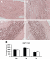
CCR5-null and wild-type (WT) infarcts exhibit comparable infiltration with macrophages. A–D: Mac2 immunohistochemistry identified macrophages in wild-type (A, C) and CCR5 KO infarcts (B, D) after 24 hours (A, B) and 72 hours of reperfusion (C, D). E: Quantitative analysis showed no significant differences in macrophage density between CCR5−/− and wild-type infarcts at all time points examined (n = 9 mice/group).
Figure 3
Figure 3
CCR5 deficiency is associated with markedly increased expression of inflammatory cytokines and chemokines in the infarcted myocardium. A: Ribonuclease protection assay analysis demonstrated that CCR5-null mice show significantly higher peak mRNA expression of the proinflammatory cytokines TNF-α, IL-1β, and IL-6 (**P < 0.01, ***P < 0.001 versus wild-type [WT]) after 6 hours of reperfusion. B: Expression of the inhibitory cytokines IL-10 and TGF-β1 was also higher in CCR5-null infarcts when compared with wild-type animals (**P < 0.01 versus wild-type). C: The IL-1β:IL-10 and IL-1β:TGF-β1 expression ratio was significantly higher in CCR5-null infarcts (**P < 0.01 versus wild-type) indicating that CCR5 absence shifted the balance of cytokine expression toward increased proinflammatory activity. D: Expression of the chemokines MIP-1α, MIP-1β, and IP-10 was also significantly higher in CCR5-null infarcts (*P < 0.05, **P < 0.01 versus wild-type) after 6 hours of reperfusion. E: MIP-2 expression was markedly higher in CCR5−/− infarcts; however, MCP-1 levels were comparable between groups (n = 8 mice/group).
Figure 4
Figure 4
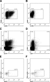
The infarcted myocardium mobilizes CCR5+ mononuclear cells. A: Flow cytometry analysis of the infarct infiltrate revealed that a significant percentage of mononuclear cells infiltrating the infarcted heart after 24 hours of reperfusion are CCR5+ (38.3%+3.7 of the CD45+ cells isolated from the infarct are CCR5+). B: As expected, CCR5 KO infarcts had no CCR5+ cells. C: In wild-type mouse infarcts a large percentage of the CCR5+ cells (38.25 ± 3.3) were identified as CD14+ monocytes. D: A significant number (3.73% ± 0.37) of the CCR5+ cells were CD4+ T cells. EF: CCR5 expression in CD4+ T cells was associated with expression of the Treg-specific marker foxp3. The CD4+/CCR5+ subset (F) had a higher percentage of foxp3+ Tregs than the CD4+/CCR5 population (E) (percentage of foxp3+ Tregs, CD4+/CCR5+ subpopulation: 45.49 ± 4.38 versus CD4+/CCR5 subpopulation: 12.47% ± 1.45, P < 0.01). The dot plots and percentages reflect a representative experiment pooling 2 infarcted hearts. Statistical analysis was based on three independent experiments using 6 infarcted hearts.
Figure 5
Figure 5
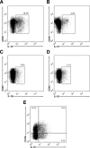
Mononuclear cells infiltrating CCR5−/− infarcts show decreased expression of IL-10. Representative flow cytometric analysis of CD45 and IL-10 expression in wild-type (A) and CCR5 KO (B) infarct mononuclear cells. Similarly, CD45 and IL-1β expression was assessed in mononuclear cell suspensions from wild-type (C) and CCR5 KO (D) infarcts. Cells isolated from CCR5 KO infarcts had reduced IL-10 expression when compared with cells from wild-type infarcts (percentage of IL-10+ cells, wild-type 19.1% ± 2.08 versus KO 5.36% ± 1.12, P < 0.01). In contrast, the percentage of IL-1β–positive cells was comparable between groups. Representative percentages indicate the fractions of CD45+ cells positive for IL-1β or IL-10. In the wild-type (E) infarcts CCR5+ subset of infarct mononuclear cells exhibited increased expression of the inhibitory cytokine IL-10 in comparison with the CCR5 subpopulation (percentage of CCR5+ cells expressing IL-10 22.48% ± 1.5 versus percentage of CCR5 cells expressing IL-10 10.34 ± 1.54, P < 0.05). Representative dot plots show expression of IL-10 in CCR5+ and CCR5 cells gated for CD45. The dot plot and percentages reflect the representative experiment pooling two hearts. Statistical analysis was based on three independent experiments using six infarcted hearts.
Figure 6
Figure 6
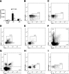
CCR5 deficiency results in impaired recruitment of regulatory T cells (Tregs) to infarcted myocardium. A: Real-time PCR showed that CCR5-null infarcts had markedly reduced mRNA expression of foxp3, a specific marker of Tregs, in comparison with WT (wild-type) infarcts after 6 hours of reperfusion (*P < 0.05 versus wild-type). B and C: Flow cytometric analysis of the CD4+ subset as a percentage of CD3+ T cells in wild-type (B) and CCR5 KO (C) infarcts. CCR5-null infarcts had a markedly lower number of CD4+ T cells when compared with wild-type animals (wild-type 45% ± 3.3 versus KO 13.3% ± 1.2, P < 0.01). D and E: Expression of CD4/foxp3 by CD3-gated mononuclear cells from wild-type (D) and CCR5 KO (E) infarcts. CCR5−/− mice had a significantly lower percentage of CD4+ foxp3+ Tregs in the infarcted heart than wild-type animals (foxp3+ CD4+ cells as a percentage of total CD3+ cells, wild-type: 13.3% ± 1.1 versus KO: 7.2% ± 1.1, P < 0.05). A representative experiment derived from isolation of mononuclear cells from two infarcted hearts for each group is shown. For statistical analysis three independent experiments (each one using pooled cells from two infarcted hearts) were performed. Flow cytometry analysis of CCR5/foxp3 (F) and IL-10/foxp3 (G) expression in wild-type splenocytes showed that foxp3+ Tregs were the predominant IL-10–producing cells (IL-10 expression was found in 27.99% ± 3.63 of foxp3+ T cells versus 0.61 ± 0.32 of foxp3- cells, P < 0.01). H and I: CCR5+ Tregs had significantly higher IL-10 expression than CCR5- Tregs (IL-10–positive cells, CCR5+/foxp3+ 42.73% ± 4.07 versus CCR5/foxp3+ 28.8 ± 4.25, P < 0.05) suggesting potent anti-inflammatory properties of the CCR5-expressing Treg subpopulation. A single representative dot plot derived from experiment using one spleen. Three independent experiments (three wild-type and three KO spleen) were used for statistical analysis.
Figure 7
Figure 7
CCR5 absence is associated with increased MMP expression in the infarcted myocardium accentuated postinfarction dilative remodeling. Compared with wild-type (WT) (A), CCR5-null animals (B) exhibited accentuated dilative remodeling after 7 days of reperfusion. Quantitative morphometry (A, B, D, E, F) and echocardiography (C, G) were used to assess remodeling-associated parameters. Echocardiographic analysis demonstrated that LVEDD increased significantly after 7 days of reperfusion in both wild-type and CCR5-null mice (**P < 0.01 versus corresponding preinfarction values). CCR5-null hearts showed increased LVEDD (C) and LVEDV (D) after 7 days of reperfusion, suggesting accentuated dilative remodeling (***P < 0.05 versus corresponding wild-type). E: Increased dilative remodeling in CCR5-null infarcts was not attributable to a difference in the size of the infarct; scar size was comparable between wild-type and CCR5-null mice after 7 days of reperfusion. F: A trend toward increased LV mass was noted in CCR5 KO mice after 7 days of reperfusion (P = 0.08), suggesting enhanced hypertrophic remodeling. G: FS significantly decreased in both wild-type and CCR5−/− mice after 7 days of reperfusion (**P < 0.01 versus corresponding preinfarction values), indicating the development of systolic dysfunction following myocardial infarction. However, the difference in fractional shortening between wild-type and CCR5 KO animals did not reach statistical significance (n = 12 mice/group). H: CCR5 KO mice had significantly higher MMP-8 and MMP-9 mRNA levels than wild-type animals after 24 hours of reperfusion (*P < 0.05, **P < 0.01 versus wild-type). In contrast, MMP-3 mRNA expression was comparable between groups. I: There was a trend toward increased MMP-2 mRNA expression in infarcted CCR5 KO hearts (P = 0.20). TIMP-1 mRNA levels, on the other hand, were significantly lower in wild-type infarcts. J: The ratios MMP-2:TIMP1, MMP-3:TIMP-1, and MMP-9:TIMP-1 were significantly higher in CCR5 KO infarcts (**P < 0.01 versus corresponding wild-type), indicating that CCR5 absence was associated with increased expression of matrix-degrading proteases in the infarcted heart (wild-type n = 9 mice, KO n = 10 mice). K: MMP-2 and MMP-9 activity were assessed in the infarcted heart using gelatin zymography. Quantitative analysis demonstrated that CCR5−/− infarcts had markedly higher active MMP-9 levels after 72 hours of reperfusion (*P < 0.05) in comparison with wild-type infarcts. In addition, active MMP-2 levels were modestly, but significantly, higher in infarcted CCR5-null hearts (*P < 0.05). L: Representative gelatin zymography experiments illustrate the increased MMP-2 and MMP-9 activity in CCR5 KO infarcts. Two control wild-type hearts, four wild-type infarcts, and four CCR5-null infarcts are shown for comparison. The arrows show the bands corresponding to active MMP-2 (a-MMP-2), latent MMP-2 (l-MMP-2), active MMP-9 (a-MMP-9), and latent MMP-9 (l-MMP-9).

References

    1. Middleton J, Neil S, Wintle J, Clark-Lewis I, Moore H, Lam C, Auer M, Hub E, Rot A. Transcytosis and surface presentation of IL-8 by venular endothelial cells. Cell. 1997;91:385–395. - PubMed
    1. Geissmann F, Jung S, Littman DR. Blood monocytes consist of two principal subsets with distinct migratory properties. Immunity. 2003;19:71–82. - PubMed
    1. Tacke F, Alvarez D, Kaplan TJ, Jakubzick C, Spanbroek R, Llodra J, Garin A, Liu J, Mack M, van Rooijen N, Lira SA, Habenicht AJ, Randolph GJ. Monocyte subsets differentially employ CCR2, CCR5, and CX3CR1 to accumulate within atherosclerotic plaques. J Clin Invest. 2007;117:185–194. - PMC - PubMed
    1. Bromley SK, Mempel TR, Luster AD. Orchestrating the orchestrators: chemokines in control of T cell traffic. Nat Immunol. 2008;9:970–980. - PubMed
    1. Nahrendorf M, Swirski FK, Aikawa E, Stangenberg L, Wurdinger T, Figueiredo JL, Libby P, Weissleder R, Pittet MJ. The healing myocardium sequentially mobilizes two monocyte subsets with divergent and complementary functions. J Exp Med. 2007;204:3037–3047. - PMC - PubMed

Publication types

MeSH terms