. 2010 May;176(5):2477-89.
doi: 10.2353/ajpath.2010.090049. Epub 2010 Apr 9.

LC3A-positive light microscopy detected patterns of autophagy and prognosis in operable breast carcinomas

Affiliations

LC3A-positive light microscopy detected patterns of autophagy and prognosis in operable breast carcinomas

Efthimios Sivridis et al. Am J Pathol. 2010 May.

Abstract

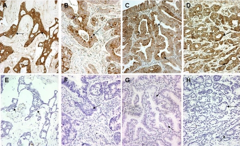
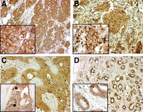
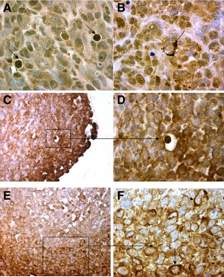
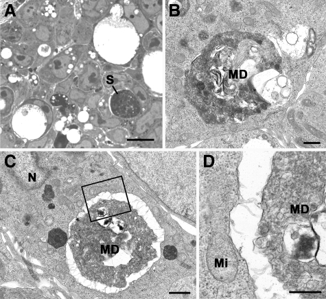
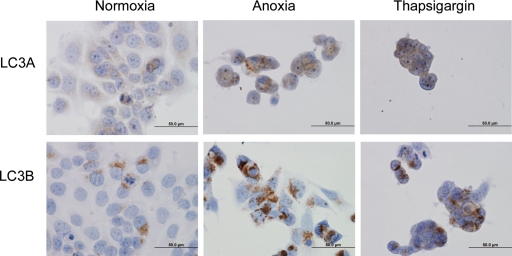
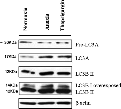
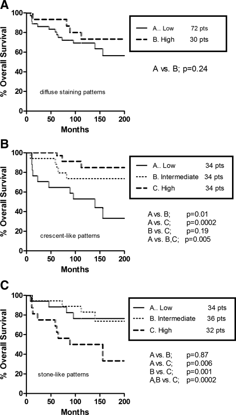
Autophagy is a self-degradation mechanism by which cells recycle their own cytoplasmic constituents and dispose of excess or defective organelles after starvation and oxygen deprivation. An antibody to the microtubule-associated protein 1 light chain 3 (LC3A), recognizing both the soluble (LC3A-I) and the membrane-bound form (LC3A-II) of the protein, was used to detect autophagic activity in 102 breast carcinomas. Three distinct patterns were recognized: (1) diffuse cytoplasmic, (2) cytoplasmic/juxta-nuclear, and (3) "stone-like" pattern--dense, rounded, amorphous structures, 5 microm on average, typically enclosed within cytoplasmic vacuoles. The diffuse cytoplasmic pattern showed a direct association with estrogen and progesterone receptor expression. The juxta-nuclear pattern indicated a similar association with hormone receptors, an inverse association with tumor size, and a favorable prognosis. By contrast, an increased number of stone-like structures, probably representing an excessive autophagic response, was related to high-grade tumors and a less favorable outcome. Interestingly, 60 additional epithelial tumors of nonbreast origin disclosed identical autophagic patterns, and so did MDA231 breast cancer xenografts and HCT116 colon tumor spheroids (also analyzed by electron microscopy). Moreover, MCF-7 human breast cancer cell lines confirmed induction of LC3A by anoxia and Thapsigargin. It is concluded that autophagy can be readily recognized in breast carcinomas by light microscopy, after immunohistochemical staining with LC3A, but the significance of the various patterns expressed would need further evaluation.


Figures

Figure 1
Figure 1
Autophagy in human tissues. Cytoplasmic, perinuclear, and stone-like patterns in malignant breast (A), colonic (B), endometrial (C), and prostatic (D) tissues stained for LC3A. The corresponding negative control sections (E–H) in the preparation of which the LC3A antibody had been replaced by normal rabbit immunoglobulin-G (Magnification ×200). Arrows in B, C, and D show stone-like structures and perinuclear staining of LC3A, whereas those in F, G, and H indicate the corresponding areas in control sections.
Figure 2
Figure 2
Patterns of LC3A expression in malignant breast tissues and the adjacent “normal” breast epithelium (magnification: large figures ×200, small figures ×1000). A: Diffuse cytoplasmic expression of LC3A in breast cancer cells (arrows). B: Juxta-nuclear pattern of LC3A expression in breast cancer cells (arrows). C: Stone-like structures within autophagic vacuoles (arrows) occupying almost the entire cytoplasm of breast cancer cells and pushing the nuclei toward the periphery. D: Nonmalignant “normal” breast glands in the proximity of the tumor with a mixed diffuse cytoplasmic and juxta-nuclear (arrows) LC3A expression.
Figure 3
Figure 3
Immunohistochemical figures from MDA231 human breast tumor xenografts (A and B: magnification ×400) and HCT116 colon tumor spheroids (C and D: magnification ×200, and E and F: magnification ×400) showing the three distinct patterns of LC3A expression (arrows).
Figure 4
Figure 4
Light (A) and electron (BD) micrographs of a section through a HCT116 tumor spheroid. A: Section showing tightly packed cells, many of which contain lucent vacuoles of various sizes. Note the cell with a large dense rounded material representing a stone-like structure (S). Scale bar is 10 μm. Plastic section/Azure A stained. B: Electron micrograph showing a small vacuole containing a mass of membranous debris (MD) within a tightly fitting vacuole in the cell cytoplasm. Scale bar is 0.5 μm. C: Low-power electron micrograph of a stone-like structure similar to that in A showing it to consist of a large mass of degenerating material (MD) within the cell cytoplasm. N indicates nucleus. Scale bar is 1 μm. D: Detail of the enclosed area in C showing the central mass to consist of a mass of MD slightly separated from the vacuolar membrane. Mi indicates mitochondrion. Scale bar is 0.5 μm.
Figure 5
Figure 5
Staining of LC3A and LC3B in MCF7 breast cancer cells treated: under normoxia, under anoxia for 48 hours, with Thapsigargin (300 nmol/L) for 24 hours.
Figure 6
Figure 6
Induction of LC3A and LC3B in MCF-7 breast cancer cells. MCF- 7 cells were treated: under anoxia for 48 hours, with Thapsigargin (300 nmol/L) and incubated under normoxic conditions for 24 hours. Levels of protein expression were measured by immunoblot analysis using antibodies against LC3A, LC3B, and β actin.
Figure 7
Figure 7
Seventeen-year cancer-specific survival rates in patients with operable breast carcinomas: diffuse cytoplasmic pattern (A), juxta-nuclear (crescentic/perinuclear) pattern (B), and “stone-like” pattern (C).

References

    1. Giatromanolaki A, Sivridis E, Koukourakis MI. Tumour angiogenesis: vascular growth and survival. APMIS. 2004;112:431–440. - PubMed
    1. Jain RK. Molecular regulation of vessel maturation. Nat Med. 2003;9:685–693. - PubMed
    1. Vaupel P, Thews O, Hoeckel M. Treatment resistance of solid tumors: role of hypoxia and anemia. Med Oncol. 2001;18:243–259. - PubMed
    1. Harris AL. Hypoxia—a key regulatory factor in tumour growth. Nat Rev Cancer. 2002;2:38–47. - PubMed
    1. Izuishi K, Kato K, Ogura T, Kinoshita T, Esumi H. Remarkable tolerance of tumor cells to nutrient deprivation: possible new biochemical target for cancer therapy. Cancer Res. 2000;60:6201–6207. - PubMed

Publication types

Substances