. 2010 Jun;176(6):2986-96.
doi: 10.2353/ajpath.2010.090665. Epub 2010 Apr 9.

TMPRSS2, a serine protease expressed in the prostate on the apical surface of luminal epithelial cells and released into semen in prostasomes, is misregulated in prostate cancer cells

Affiliations

TMPRSS2, a serine protease expressed in the prostate on the apical surface of luminal epithelial cells and released into semen in prostasomes, is misregulated in prostate cancer cells

Ya-Wen Chen et al. Am J Pathol. 2010 Jun.

Abstract

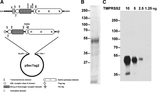
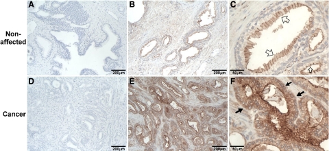
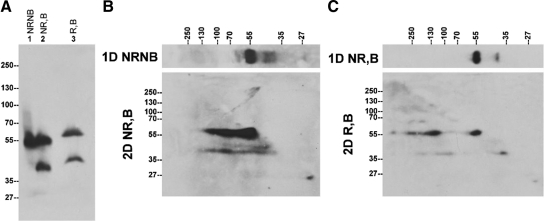
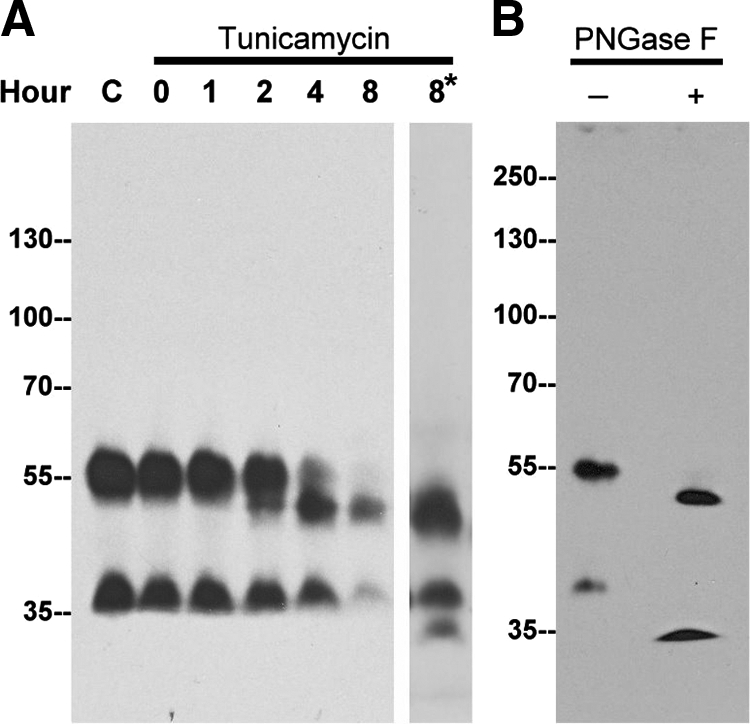
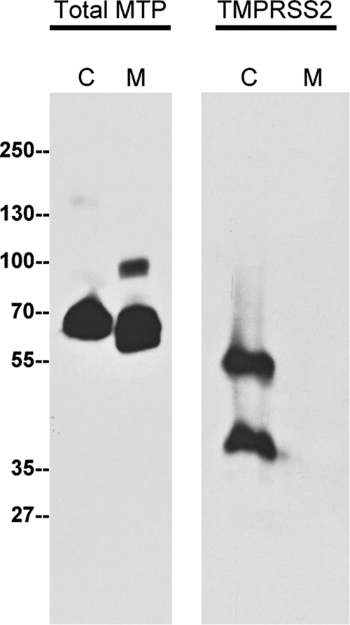
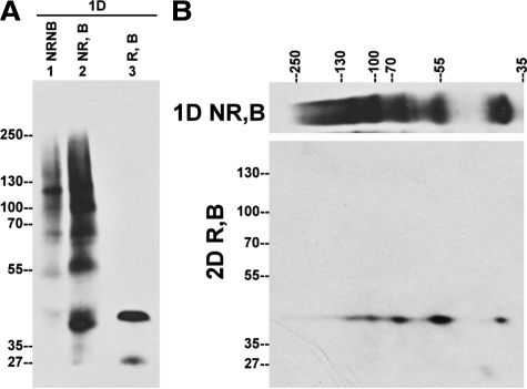
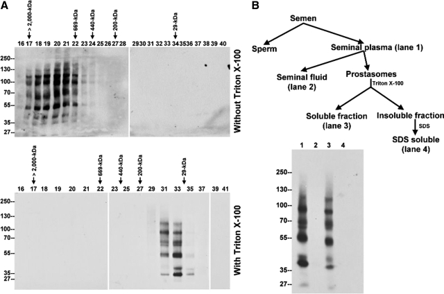
TMPRSS2, a type II transmembrane serine protease, is highly expressed by the epithelium of the human prostate gland. To explore the regulation and function of TMPRSS2 in the prostate, a panel of monoclonal antibodies with high sensitivity and specificity were generated. Immunodetection showed TMPRSS2 on the apical plasma membrane of the prostate luminal cells and demonstrated its release into semen as a component of prostasomes, organelle-like vesicles that may facilitate sperm function and enhance male reproduction. In prostate cancer cells, TMPRSS2 expression was increased and the protein mislocalized over the entire tumor cell membrane. In both LNCaP prostate cancer cells and human semen, TMPRSS2 protein was detected predominantly as inactive zymogen forms as part of an array of multiple noncovalent and disulfide-linked complexes, suggesting that TMPRSS2 activity may be regulated by unconventional mechanisms. Our data suggested that TMPRSS2, an apical surface serine protease, may have a normal role in male reproduction as a component of prostasomes. The aberrant cellular localization, and increased expression of the protease seen in cancer, may contribute to prostate tumorigenesis by providing access of the enzyme to nonphysiological substrates and binding-proteins.


Figures

Figure 1
Figure 1
TMPRSS2 recombinant protein and mAbs. A: Expression of TMPRSS2 in mammalian cells. A schematic representation of the structure of TMPRSS2 is presented at the top. The protease consists of 492 amino acids, has a calculated mass of 53,859 Da, and contains a transmembrane domain, a scavenger receptor cysteine-rich (SRCR) domain, a LDLRA domain, and a serine protease domain. The two putative N-glycosylation sites are at N-213 and N-249. The serine protease domain is at the carboxyl terminus, and the active site triad was labeled as H-D-S. A truncated and mutated TMPRSS2 expression construct was generated as described in the Materials and Methods section resulting in the construct shown. B: Purification of TMPRSS2. The recombinant TMPRSS2 protein was produced and purified from HEK 293T cells and resolved by SDS-PAGE and stained by colloidal Coomassie blue to show the protein patterns and to asses the purity of the TMPRSS2 preparation. C: Characterization of a TMPRSS2 monoclonal antibody. To evaluate the sensitivity of the TMPRSS2 monoclonal antibody, AL20, different amounts of purified TMPRSS2 protein as indicated were resolved by SDS-PAGE under reducing and boiled conditions. Immunoblot analysis was conducted with the AL20 monoclonal antibody at 2 μg/ml.
Figure 2
Figure 2
Cellular localization of TMPRSS2 in normal and carcinomatous prostate tissues. Paraffin-embedded human prostate tumor tissue sections were stained by immunohistochemistry using TMPRSS2 mAb AL20 (B, C, E, and F) and mouse IgG (A and D). Positive staining for TMPRSS2 was observed as brown precipitates (diaminobenzidine), and the nuclei were counterstained with hematoxylin. The micrographs in AC were taken from noncancerous acini portions of the prostate tumor (nonaffected) and DF from the cancer portions. The sizes of bars are indicated in each panel. Open arrows show TMPRSS2 on the apical surface of luminal cells. Closed arrows show the difference of TMPRSS2 protein expression in nonaffected prostate tissue and carcinomatous prostate tissue.
Figure 3
Figure 3
The expression status of endogenous TMPRSS2 protein in human prostate cancer cells. A: TMPRSS2 protein expression in LNCaP prostate cancer cells. LNCaP cells were lysed using PBS containing 1% Triton X-100. Equal amounts of total cellular proteins were mixed with SDS sample buffer in the absence (NR) or presence (R) of reducing agents and incubated either at room temperature (NB) or 95°C (B) for 5 minutes, followed by SDS-PAGE and Western blot analysis using the monoclonal antibody AL20. NRNB indicates nonreducing and nonboiled conditions; NR,B, nonreducing and boiled conditions; R,B, reducing and boiled conditions. B: TMPRSS2 is present in non-covalent-linked complexes in LNCaP cells. LNCaP cell lysates were resolved by diagonal gel electrophoresis under nonreducing and nonboiled conditions in the first dimension and under nonreducing and boiled condition in the second dimension. Immunoblot analysis of the second dimensional gel was performed with the TMPRSS2 mAb AL20. C: TMPRSS2 is also present in disulfide bond-linked complexes in LNCaP cells. LNCaP cell lysates were resolved by diagonal gel electrophoresis under nonreducing and boiled conditions in the first dimension and under reducing and boiled condition in the second dimension. Immunoblot analysis of the second dimensional gel was performed with the TMPRSS2 mAb AL20.
Figure 4
Figure 4
N-glycosylation of TMPRSS2 is responsible for a modest increase in molecular mass. A: LNCaP cells were treated with tunicamycin (5 μg/ml) for the indicated times. Cell lysates were analyzed for TMPRSS2 by immunoblot using the mAb AL20. To facilitate detection of the deglycosylated 40-kDa species, a longer exposure of the immunoblot for the 8-hour time point was taken (8*). B: LNCaP cell lysates were incubated in the absence (−) or presence (+) of PNGase F, and TMPRSS2 was probed with the mAb AL20.
Figure 5
Figure 5
Shedding of TMPRSS2 to the extracellular milieu by LNCaP cells is very limited. LNCaP cells were grown in serum-free media for 24 hours, and then the conditioned medium and cell lysates were harvested. The conditioned medium was concentrated up to 100 fold. The cell lysates (C) and the conditioned medium (M) were analyzed for total matriptase (Total MTP) using the matriptase mAb M32 and TMPRSS2 using the mAb AL20.
Figure 6
Figure 6
TMPRSS2 is present in multiple complexes in human semen. A: Equal amount of human semen were mixed with SDS sample buffer in the absence (NR) or presence (R) of reducing agents and incubated either at room temperature (NB) or 95°C (B) for 5 minutes, followed by SDS-PAGE and Western blot analysis using TMPRSS2 mAb AL20. B: Human semen was resolved by diagonal gel electrophoresis under nonreducing and boiled conditions in the first dimension and under reducing and boiled conditions in the second dimension. Immunoblot analysis of the second dimensional gel was performed with the TMPRSS2 mAb AL20.
Figure 7
Figure 7
TMPRSS2 is expressed as components of the prostasome. A: Seminal plasma was fractionated in the absence (top) or presence (bottom) of 1% Triton X-100 by the Superose 6 sizing column. The Superose 6 fractions were examined by immunoblot analysis using TMPRSS2 mAb AL20. In the absence of Triton X-100, TMPRSS2 was detected in the void volume as indicated by co-elution with blue dextran (top). In the presence of nonionic detergent, TMPRSS2 was eluted in the fractions whose apparent molecular masses were distributed from 200 kDa to 29 kDa as determined by comparison with standard proteins which included blue dextran (>2000 kDa), thyroglobulin (669 kDa), apoferritin (443 kDa), b-amylase (200 kDa), carbonic anhydrase (29 kDa) as indicated (bottom). B: Top: Flow chart of human semen fractionation. Human semen was fractionated by centrifugation at 1,000g for 15 minutes into sperm fractions and seminal plasma. The latter was further fractionated by centrifugation at 105,000g for 2 hours into the insoluble fractions, representing the prostasomes, and the soluble fractions, the seminal fluids. Prostasomes were then lysed with 1% Triton X-100 in PBS, and the insoluble fractions were further extracted by 1% SDS in PBS. Bottom: The four semen fractions were subjected to SDS-PAGE and followed by immunoblot analyses for TMPRSS2. The TTSP, detected in seminal plasma (lane 1), was fractionated into the prostasome Triton X-100–soluble fractions (lane 3), but not in the seminal fluid (lane 2) and the Triton X-100–insoluble fractions (lane 4).

References

    1. Paoloni-Giacobino A, Chen H, Peitsch MC, Rossier C, Antonarakis SE. Cloning of the TMPRSS2 gene, which encodes a novel serine protease with transmembrane. LDLRA, and SRCR domains and maps to 21q223. Genomics. 1997;44:309–320. - PubMed
    1. Lin B, Ferguson C, White JT, Wang S, Vessella R, True LD, Hood L, Nelson PS. Prostate-localized and androgen-regulated expression of the membrane-bound serine protease TMPRSS2. Cancer Res. 1999;59:4180–4184. - PubMed
    1. Jacquinet E, Rao NV, Rao GV, Zhengming W, Albertine KH, Hoidal JR. Cloning and characterization of the cDNA and gene for human epitheliasin. Eur J Biochem. 2001;268:2687–2699. - PubMed
    1. Hooper JD, Clements JA, Quigley JP, Antalis TM. Type II transmembrane serine proteases. Insights into an emerging class of cell surface proteolytic enzymes. J Biol Chem. 2001;276:857–860. - PubMed
    1. Netzel-Arnett S, Hooper JD, Szabo R, Madison EL, Quigley JP, Bugge TH, Antalis TM. Membrane anchored serine proteases: a rapidly expanding group of cell surface proteolytic enzymes with potential roles in cancer. Cancer Metastasis Rev. 2003;22:237–258. - PubMed

Publication types

MeSH terms