Review
. 2010 Jun 18;285(25):18979-83.
doi: 10.1074/jbc.R109.022939. Epub 2010 Apr 9.

Timing, coordination, and rhythm: acrobatics at the DNA replication fork

Affiliations
Review

Timing, coordination, and rhythm: acrobatics at the DNA replication fork

Samir M Hamdan et al. J Biol Chem. .

Abstract

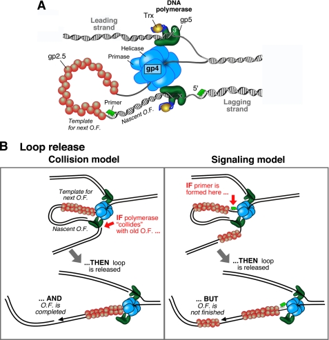
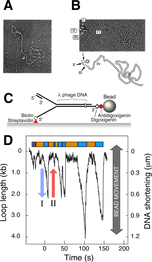
In DNA replication, the antiparallel nature of the parental duplex imposes certain constraints on the activity of the DNA polymerases that synthesize new DNA. The leading-strand polymerase advances in a continuous fashion, but the lagging-strand polymerase is forced to restart at short intervals. In several prokaryotic systems studied so far, this problem is solved by the formation of a loop in the lagging strand of the replication fork to reorient the lagging-strand DNA polymerase so that it advances in parallel with the leading-strand polymerase. The replication loop grows and shrinks during each cycle of Okazaki fragment synthesis. The timing of Okazaki fragment synthesis and loop formation is determined by a subtle interplay of enzymatic activities at the fork. Recent developments in single-molecule techniques have enabled the direct observation of these processes and have greatly contributed to a better understanding of the dynamic nature of the replication fork. Here, we will review recent experimental advances, present the current models, and discuss some of the exciting developments in the field.


Figures

FIGURE 1.
FIGURE 1.
A, organization of the bacteriophage T7 replication fork. gp4 encircles the lagging strand and mediates both the unwinding of dsDNA via its helicase domain and the synthesis of RNA primers via the primase domain. The T7 DNA polymerases are stably bound to gp4 and incorporate nucleotides on the leading and lagging strands. The DNA polymerase is a 1:1 complex of T7 gp5 and E. coli Trx. The ssDNA extruded behind the helicase is coated by the ssDNA-binding protein gp2.5. A replication loop is formed in the lagging strand to align it with the leading strand. The lagging-strand DNA polymerase initiates Okazaki fragment (O.F.) synthesis using RNA primers (green segments). B, schematic depiction of the two models that describe replication loop release. In the collision model (left panel), the replication loop is released when the lagging-strand polymerase collides with the 5′ terminus of the previous Okazaki fragment. In the signaling model (right panel), the synthesis of a new primer triggers the release of the replication loop prior to the completion of the nascent Okazaki fragment.
FIGURE 2.
FIGURE 2.
A, observation of replication loops by EM. Coordinated DNA synthesis is carried out on a 70-bp minicircle substrate. The replication proteins appear as a globular object. The dsDNA loop extending from the replisome represents the nascent Okazaki fragment. Both the small minicircle and the condensed gp2.5-ssDNA complex are obscured by the protein mass containing the replisome. The figure is from Ref. . B, observation of Okazaki fragments by EM. The replication reaction was carried out as described for A, deproteinated, and treated with SSB. SSB extends ssDNA and enables its visualization as a DNA segment with increased diameter. Segment I indicates the template for the next Okazaki fragment produced by the helicase, segment II is the nascent Okazaki fragment, segment III is the template of the nascent Okazaki fragment, and segment IV is the long dsDNA of previously synthesized Okazaki fragments. The figure is from Ref. . C, flow-stretching individual DNA molecules. Duplex λ-DNA (48.5 kb) is modified to contain a replication fork. The lagging strand of the forked end is coupled to the surface. The other end of the DNA is attached to a bead. A constant laminar flow applies a well controlled drag force to the bead and stretches the DNA molecule. The length of the individual DNA molecules is measured by imaging the beads and tracking their positions. The figure is from Ref. . D, dynamic single-molecule observation of replication loops produced by individual replisomes. The trajectory shows a time course of the length of a single DNA molecule during replication. The DNA shortening corresponds to loop growth during leading- and lagging-strand synthesis (blue arrow) and is followed by a rapid length increase when a loop is released (red arrow). The lag phases between looping events and the loop growth phases are shown as orange and cyan boxes, respectively. The figure is from Ref. .

References

    1. Benkovic S. J., Valentine A. M., Salinas F. (2001) Annu. Rev. Biochem. 70, 181–208 - PubMed
    1. Johnson A., O'Donnell M. (2005) Annu. Rev. Biochem. 74, 283–315 - PubMed
    1. Hamdan S. M., Richardson C. C. (2009) Annu. Rev. Biochem. 78, 205–243 - PubMed
    1. Ogawa T., Okazaki T. (1980) Annu. Rev. Biochem. 49, 421–457 - PubMed
    1. Tabor S., Huber H. E., Richardson C. C. (1987) J. Biol. Chem. 262, 16212–16223 - PubMed

Publication types

LinkOut - more resources