. 2010 Jun;192(12):3144-58.
doi: 10.1128/JB.00031-10. Epub 2010 Apr 9.

RmpA regulation of capsular polysaccharide biosynthesis in Klebsiella pneumoniae CG43

Affiliations

RmpA regulation of capsular polysaccharide biosynthesis in Klebsiella pneumoniae CG43

H Y Cheng et al. J Bacteriol. 2010 Jun.

Abstract

Sequence analysis of the large virulence plasmid pLVPK in Klebsiella pneumoniae CG43 revealed the presence of another mucoid factor encoding gene rmpA besides rmpA2. Promoter activity measurement indicated that the deletion of rmpA reduced K2 capsular polysaccharide (CPS) biosynthesis, resulting in decreased colony mucoidy and virulence in mice. Introduction of a multicopy plasmid carrying rmpA restored CPS production in the rmpA or rmpA2 mutant but not in the rcsB mutant. Transformation of the rmpA deletion mutant with an rcsB-carrying plasmid also failed to enhance CPS production, suggesting that a cooperation of RmpA with RcsB is required for regulatory activity. This was further corroborated by the demonstration of in vivo interaction between RmpA and RcsB using two-hybrid analysis and coimmunoprecipitation analysis. A putative Fur binding box was only found at the 5' noncoding region of rmpA. The promoter activity analysis indicated that the deletion of fur increased the rmpA promoter activity. Using electrophoretic mobility shift assay, we further demonstrated that Fur exerts its regulatory activity by binding directly to the promoter. As a result, the fur deletion mutant exhibited an increase in colony mucoidy, CPS production, and virulence in mice. In summary, our results suggested that RmpA activates CPS biosynthesis in K. pneumoniae CG43 via an RcsB-dependent manner. The expression of rmpA is regulated by the availability of iron and is negatively controlled by Fur.


Figures

FIG. 1.
FIG. 1.
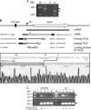
Comparison of rmpA and rmpA2 containing PAI-like regions and demonstration of rmpA expression. (A) The arrows indicate predicted open reading frames and insertion sequences. The reporter constructs used for promoter activity measurement are shown below. (B) Assessment of rmpA expression by PCR and Southern hybridization analysis. In the upper panel, the templates for the PCR include a plasmid carrying rmpA gene (lane C), the extracted total RNA or reverse-transcribed cDNA from K. pneumoniae CG43S3 (wild type) or rmpA2 deletion mutant (ΔrmpA2). The lower panel shows the result of Southern blot analysis of the same gel with a probe specific to the rmpA gene; the arrow indicates the expected size of the PCR product.
FIG. 2.
FIG. 2.
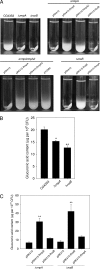
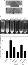
Comparison of precipitation speeds and K2 CPS production in K. pneumoniae strains. (A) The strains tested were grown overnight in LB broth at 37°C and subjected to centrifugation at 4,000 × g for 5 min. (B) The glucuronic acid content, which served as an indicator of K2 CPS, was determined from overnight K. pneumoniae cultures. The results are expressed as the average of the triplicate samples. Error bars indicate standard deviations. *, P < 0.01; **, P < 0.001 compared to the parental strain CG43S3 (n ≥ 3). (C) Quantification of Klebsiella K2 CPS production. The results are expressed as an average of triplicate samples. Error bars indicate standard deviations. **, P < 0.001 compared to the same strain carrying pRK415 (n ≥ 3).
FIG. 3.
FIG. 3.
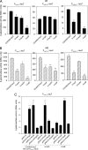
Expression of K2 cps genes in various genetic backgrounds. The β-galactosidase activities of K2 cps Porf1-2::lacZ, Porf3-15::lacZ, and Porf16-17::lacZ in K. pneumoniae CG43S3ΔlacZ (wild-type) and its isogenic strains (ΔrmpAΔlacZ, ΔrmpA2ΔlacZ, and ΔrcsBΔlacZ) harboring each of the reporter plasmids pOrf12, pOrf315, or pOrf1617 were determined from log-phase cultures grown in LB broth (A) or M9-glucose medium (B). The results are shown as an average of triplicate samples. Error bars indicate standard deviations. *, P < 0.01; **, P < 0.001 compared to the parental strain CG43S3ΔlacZ (n ≥ 3). (C) The K. pneumoniae CG43S3ΔlacZ (wild-type) and its isogenic strains (ΔrmpAΔlacZ and ΔrcsBΔlacZ), each carrying a chromosomally integrated K2 cps Porf1-2::lacZ cassette, were transformed individually with pRK415 and its derived plasmids. The β-galactosidase activities were determined from log-phase (OD600 of 0.7) cultures grown in LB broth. The results are shown as the average of the triplicate samples. Error bars indicate standard deviations. **, P < 0.001 compared to each strain carrying pRK415 (n ≥ 3).
FIG. 4.
FIG. 4.
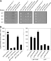
Bacterial two-hybrid analysis of the interaction between RcsA/RcsB, RcsB/RmpA, and RcsB/RmpA2 proteins. (A) The growth of serially diluted cultures of E. coli reporter strains cotransformed with pTRG and pBT or the derived plasmids was investigated on the indicator plate. (B) The E. coli reporter strains cotransformed with pTRG and pBT or the derived plasmids were grown to log phase (OD600 of 0.5) in LB broth and induced with IPTG, and the β-galactosidase activities were determined. The results are shown as the average of the triplicate samples. Error bars indicate standard deviations. *, P < 0.0001 compared to negative control strain carrying the bait vector pBT and the target vector pTRG (n ≥ 3). **, P < 0.001 compared to the strain carrying the bait vector pBT (n ≥ 3).
FIG. 5.
FIG. 5.
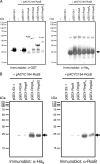
Coimmunoprecipitation analysis of the interaction between RcsA/RcsB, RcsB/RmpA, and RcsB/RmpA2 proteins. (A) Results of immunoblot analysis of Pre-IP samples using anti-GST (α-GST) or anti-His6 (α-His6) monoclonal antibodies showing, respectively, the expression of GST fusion proteins and RcsB-His6. Samples were supernatants of induced bacterial cell lysates prepared from E. coli BL21(DE3) with or without (-) different combinations of expression vectors as indicated above the figure, and 10 μg of total protein was loaded in each well. The asterisks indicate the expected size of GST and GST fusion proteins. The arrow indicates the expected size of RcsB-His6. (B) Results of immunoblot analysis of IP samples showing the interaction between the recombinant proteins. Protein complexes were precipitated with glutathione-Sepharose beads, separated by SDS-PAGE, and immunoblotted with anti-His6 (α-His6) monoclonal antibody or anti-RcsB-His6 (α-RcsB) polyclonal antiserum. The arrows indicate the expected size of RcsB-His6.
FIG. 6.
FIG. 6.
Effect of fur deletion or iron depletion on the activity of PrmpA::lacZ, PrmpA2::lacZ, PiucA::lacZ, and PiroB::lacZ. The β-galactosidase activities of K. pneumoniae CG43S3ΔlacZ (wild type) or its isogenic fur deletion mutant (Δfur) carrying, respectively, placZ15-PrmpA (PrmpA::lacZ), placZ15-PrmpA2 (PrmpA2::lacZ), placZ15-PiucA (PiucA::lacZ), or placZ15-PiroB (PiroB::lacZ) were determined from cultures grown in LB (A) or M9-glucose (B) medium. The media were supplemented with (white bars) or without (black bars and striped bars) 0.2 mM iron chelator 2,2-dipyridyl (+Dip). The results are shown as the average of the triplicate samples. Error bars indicate the standard deviations. *, P < 0.01; **, P < 0.001 compared to the parental strain CG43S3ΔlacZ grown in media without supplements (n ≥ 3).
FIG. 7.
FIG. 7.
EMSA of the recombinant His6-Fur and its target promoters. (A) Diagrammatic representation of the iucA, iroB, and rmpA loci. The large arrows represent the open reading frames. The relative positions of the primer sets used in PCR amplification of the DNA probes are, respectively, indicated, and the numbers denote the relative positions to the translational start site. Names and sizes of the DNA probes are shown on the left. The dashed boxes indicate the predicted Fur binding sequences, and the alignment result is shown below. (B) Binding of His6-Fur to its target promoters. The 32P-labeled DNA probes of PiucA (P1), PiroB (P2), and PrmpA (P3) were incubated with increasing amounts of recombinant His6-Fur protein as indicated. (C) Binding of His6-Fur to PrmpA. The 32P-labeled DNA probes of PrmpA (P3, P4, P5, and P6) were incubated with increasing amounts of recombinant His6-Fur protein as indicated. (D) Binding specificity of His6-Fur to PrmpA. The 32P-labeled DNA probe of PrmpA (P3) was incubated with various amounts of His6-Fur as indicated. Binding specificity was investigated by adding indicated amounts of unlabeled specific (P3 and P5, lanes 5 to 7 and lane 12) or nonspecific (pT7-7, pUC19 plasmid DNA, rmpA gene, and P6, lanes 8 to 11) competitor DNA fragments.
FIG. 8.
FIG. 8.
Identification of rmpA transcription start site by 5′-RACE. (A) Electrophoresis of the 5′-RACE PCR products. M, DNA ladder. The templates used in each PCR include the cDNA from K. pneumoniae CG43S3 (primary PCR) (lane 1), reverse transcription reaction mixture without transcriptase as a negative control (lane 2), or 100-fold-diluted primary PCR mixture (nested PCR) (lane 3). The arrows indicate the expected sizes of the PCR products. (B) Schematic representation of the rmpA locus and the 5′-RACE experimental design. The large arrow represents the RmpA open reading frame. The relative positions of the primers and expected sizes of the products in the primary and nested PCRs are indicated. The rmpA transcriptional start site is marked as S1. The potential −10 and −35 sites and the translational start site are underlined. (C) Assessment of rmpA transcription by limiting-dilution RT-PCR. The templates used in each reaction include total RNA (lane 1) and the 4-fold serially diluted cDNA (lanes 2 to 5 represent, respectively 1-, 1/4-, 1/16-, and 1/64-fold dilutions) from K. pneumoniae CG43S3 or its isogenic fur mutant. The upper panel shows the results of electrophoresis of the RT-PCR products amplified with primers RTrmpA01/RTrmpA02. The RT-PCR products using 23S rRNA gene primers are shown in the lower panel. The expression levels of rmpA were quantified and normalized with 23S rRNA using ImageJ software (National Institutes of Health) as shown below. The rmpA expression level of the undiluted cDNA from K. pneumoniae CG43S3 was set as 100. The arrows indicate the expected size of the PCR products. M, DNA ladder.
FIG. 9.
FIG. 9.
Phenotype comparison of K. pneumoniae CG43S3, the fur deletion mutant, and the complemented strain. (A) The bacterial strains on LB agar plates incubated at 37°C for 48 h. (B) Sedimentation test and quantification of K2 CPS for strains grown overnight in LB broth at 37°C and subjected to centrifugation at 4,000 × g for 5 min. (C) The glucuronic acid contents (μg/109 CFU) determined from overnight K. pneumoniae cultures, expressed as the average of the triplicate samples ± the standard deviations. *, P < 0.001 compared to the parental strain CG43S3 (n ≥ 3). **, P < 0.001 compared to each strain carrying pRK415 (n ≥ 3).

References

    1. Arakawa, Y., R. Wacharotayankun, T. Nagatsuka, H. Ito, N. Kato, and M. Ohta. 1995. Genomic organization of the Klebsiella pneumoniae cps region responsible for serotype K2 capsular polysaccharide synthesis in the virulent strain Chedid. J. Bacteriol. 177:1788-1796. - PMC - PubMed
    1. Baichoo, N., and J. D. Helmann. 2002. Recognition of DNA by Fur: a reinterpretation of the Fur box consensus sequence. J. Bacteriol. 184:5826-5832. - PMC - PubMed
    1. Brill, J. A., C. Quinlan-Walshe, and S. Gottesman. 1988. Fine-structure mapping and identification of two regulators of capsule synthesis in Escherichia coli K-12. J. Bacteriol. 170:2599-2611. - PMC - PubMed
    1. Brisse, S., C. Fevre, V. Passet, S. Issenhuth-Jeanjean, R. Tournebize, L. Diancourt, and P. Grimont. 2009. Virulent clones of Klebsiella pneumoniae: identification and evolutionary scenario based on genomic and phenotypic characterization. PLoS One 4:e4982. - PMC - PubMed
    1. Campos, M. A., M. A. Vargas, V. Regueiro, C. M. Llompart, S. Alberti, and J. A. Bengoechea. 2004. Capsule polysaccharide mediates bacterial resistance to antimicrobial peptides. Infect. Immun. 72:7107-7114. - PMC - PubMed

Publication types

MeSH terms