Pathology and viral antigen distribution of lethal pneumonia in domestic cats due to pandemic (H1N1) 2009 influenza A virus
- PMID: 20382823
- PMCID: PMC4165647
- DOI: 10.1177/0300985810368393
Pathology and viral antigen distribution of lethal pneumonia in domestic cats due to pandemic (H1N1) 2009 influenza A virus
Abstract
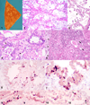
A novel swine-origin H1N1 influenza A virus has been identified as the cause of the 2009 influenza pandemic in humans. Since then, infections with the pandemic (H1N1) 2009 influenza virus have been documented in a number of animal species. The first known cases of lethal respiratory disease associated with pandemic (H1N1) 2009 influenza virus infection in house pets occurred in domestic cats in Oregon. A 10-year-old neutered domestic shorthair and an 8-year-old spayed domestic shorthair died shortly after developing severe respiratory disease. Grossly, lung lobes of both cats were diffusely firm and incompletely collapsed. Histologically, moderate to severe necrotizing to pyonecrotizing bronchointerstitial pneumonia was accompanied by serofibrinous exudation and hyaline membranes in the alveolar spaces. Influenza A virus was isolated from nasal secretions of the male cat and from lung homogenate of the female cat. Both isolates were confirmed as pandemic (H1N1) 2009 influenza virus by real-time reverse transcriptase polymerase chain reaction. With immunohistochemistry, influenza A viral antigen was demonstrated in bronchiolar epithelial cells, pneumocytes, and alveolar macrophages in pneumonic areas. The most likely sources of infection were people in the household with influenza-like illness or confirmed pandemic (H1N1) 2009 influenza. The 2 cases reported here provide, to the best of the authors' knowledge, the first description of the pathology and viral antigen distribution of lethal respiratory disease in domestic cats after natural pandemic (H1N1) 2009 influenza virus infection, probably transmitted from humans.
Figures


Similar articles
-
Acute bronchointerstitial pneumonia in two indoor cats exposed to the H1N1 influenza virus.J Vet Emerg Crit Care (San Antonio). 2014 Nov-Dec;24(6):715-23. doi: 10.1111/vec.12179. Epub 2014 Apr 8. J Vet Emerg Crit Care (San Antonio). 2014. PMID: 24712839
-
Influenza A pandemic (H1N1) 2009 virus outbreak in a cat colony in Italy.Zoonoses Public Health. 2011 Dec;58(8):573-81. doi: 10.1111/j.1863-2378.2011.01406.x. Epub 2011 Apr 11. Zoonoses Public Health. 2011. PMID: 21824359
-
Domestic cats are susceptible to infection with low pathogenic avian influenza viruses from shorebirds.Vet Pathol. 2013 Jan;50(1):39-45. doi: 10.1177/0300985812452578. Epub 2012 Jun 25. Vet Pathol. 2013. PMID: 22732359 Free PMC article.
-
Viral pathogens and acute lung injury: investigations inspired by the SARS epidemic and the 2009 H1N1 influenza pandemic.Semin Respir Crit Care Med. 2013 Aug;34(4):475-86. doi: 10.1055/s-0033-1351122. Epub 2013 Aug 11. Semin Respir Crit Care Med. 2013. PMID: 23934716 Free PMC article. Review.
-
The pathology and pathogenesis of experimental severe acute respiratory syndrome and influenza in animal models.J Comp Pathol. 2014 Jul;151(1):83-112. doi: 10.1016/j.jcpa.2014.01.004. Epub 2014 Jan 15. J Comp Pathol. 2014. PMID: 24581932 Free PMC article. Review.
Cited by
-
Genetic and serologic surveillance of canine (CIV) and equine (EIV) influenza virus in Nuevo León State, México.PeerJ. 2019 Dec 17;7:e8239. doi: 10.7717/peerj.8239. eCollection 2019. PeerJ. 2019. PMID: 31871842 Free PMC article.
-
Reverse Zoonosis of COVID-19: Lessons From the 2009 Influenza Pandemic.Vet Pathol. 2021 Mar;58(2):234-242. doi: 10.1177/0300985820979843. Epub 2020 Dec 9. Vet Pathol. 2021. PMID: 33295843 Free PMC article. Review.
-
Alveolar Macrophages in Viral Respiratory Infections: Sentinels and Saboteurs of Lung Defense.Int J Mol Sci. 2025 Jan 5;26(1):407. doi: 10.3390/ijms26010407. Int J Mol Sci. 2025. PMID: 39796262 Free PMC article. Review.
-
Serological Evidence for Influenza A Viruses Among Domestic Dogs and Cats in Kyiv, Ukraine.Vector Borne Zoonotic Dis. 2021 Jul;21(7):483-489. doi: 10.1089/vbz.2020.2746. Epub 2021 Apr 20. Vector Borne Zoonotic Dis. 2021. PMID: 33877900 Free PMC article.
-
Highly pathogenic avian influenza (HPAI) H5 virus exposure in domestic cats and rural stray cats, the Netherlands, October 2020 to June 2023.Euro Surveill. 2024 Oct;29(44):2400326. doi: 10.2807/1560-7917.ES.2024.29.44.2400326. Euro Surveill. 2024. PMID: 39484684 Free PMC article.
References
-
- Burgesser KM, Hotaling S, Schiebel A, Ashbaugh SE, Roberts SM, Collins JK. Comparison of PCR, virus isolation, and indirect fluorescent antibody staining in the detection of naturally occurring feline herpesvirus infections. J Vet Diagn Invest. 1999;11:122–126. - PubMed
-
- Caswell JL, Williams KJ. Respiratory System. In: Maxie MG, editor. Jubb, Kennedy, and Palmer’s Pathology of Domestic Animals. 5th ed. Vol. 2. Philadelphia, USA: Elsevier Saunders; 2007. pp. 523–653.
-
- Centers for Disease Control and Prevention (CDC) Swine influenza A (H1N1) infection in two children - Southern California, March–April 2009. MMWR Morb Mortal Wkly Rep. 2009;58(Dispatch):1–3. - PubMed
-
- Centers for Disease Control and Prevention (CDC) Update: Infections with a swine-origin influenza A (H1N1) virus - United States and other countries, April 28, 2009. MMWR Morb Mortal Wkly Rep. 2009;58(16):431–433. - PubMed
-
- Centers for Disease Control and Prevention (CDC) Outbreak of swine-origin influenza A (H1N1) virus infection - Mexico, March–April 2009. MMWR Morb Mortal Wkly Rep. 2009;58(17):467–470. - PubMed
Publication types
MeSH terms
Substances
Grants and funding
LinkOut - more resources
Full Text Sources
Miscellaneous

