. 2010 Jun;185(2):573-85.
doi: 10.1534/genetics.110.116293. Epub 2010 Apr 9.

Genes required for cellular UNC-6/netrin localization in Caenorhabditis elegans

Affiliations

Genes required for cellular UNC-6/netrin localization in Caenorhabditis elegans

Taro Asakura et al. Genetics. 2010 Jun.

Abstract

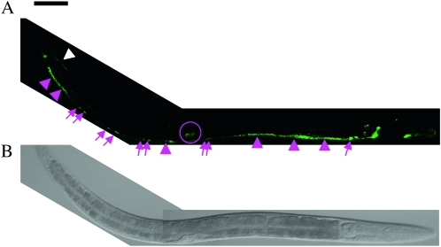
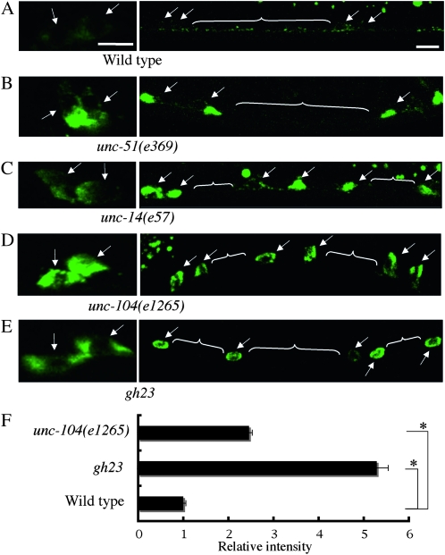
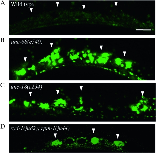
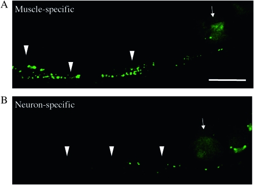
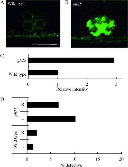
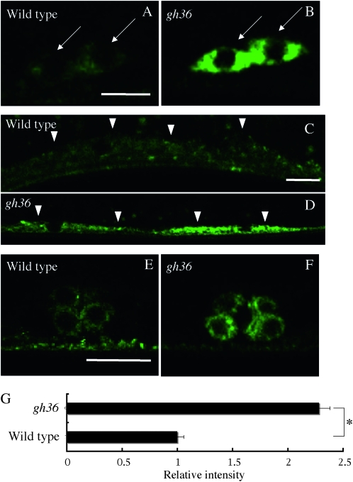
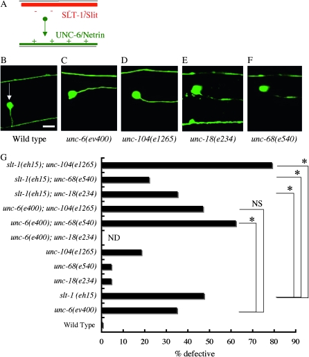
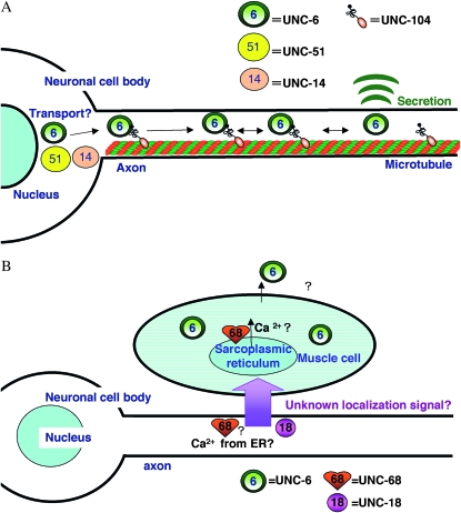
UNC-6/Netrin is an evolutionarily conserved, secretory axon guidance molecule. In Caenorhabditis elegans, UNC-6 provides positional information to the axons of developing neurons, probably by establishing a concentration gradient from the ventral to the dorsal side of the animal. Although the proper localization of UNC-6 is important for accurate neuronal network formation, little is known about how its localization is regulated. Here, to examine the localization mechanism for UNC-6, we generated C. elegans expressing UNC-6 tagged with the fluorescent protein Venus and identified 13 genes, which are involved in the cellular localization of VenusUNC-6. For example, in unc-51, unc-14, and unc-104 mutants, the neurons showed an abnormal accumulation of VenusUNC-6 in the cell body and less than normal level of VenusUNC-6 in the axon. An aberrant accumulation of VenusUNC-6 in muscle cells was seen in unc-18 and unc-68 mutants. unc-51, unc-14, and unc-104 mutants also showed defects in the guidance of dorso-ventral axons, suggesting that the abnormal localization of UNC-6 disturbed the positional information it provides. We propose that these genes regulate the process of UNC-6 secretion: expression, maturation, sorting, transport, or exocytosis. Our findings provide novel insight into the localization mechanism of the axon guidance molecule UNC-6/Netrin.


Figures

F<sc>igure</sc> 1.—
Figure 1.—
Expression of Venus∷UNC-6 in living C. elegans. An L4 worm. Right lateral view, anterior is to the right. (A) Expression of Venus∷UNC-6. (B) DIC image of the same worm. Bar, 50 μm. As described previously (Wadsworth et al. 1996), ventral neurons expressed Venus∷UNC-6 (magenta arrows). In addition, ventral muscle (magenta arrowheads), dorsal muscle (white arrowheads), and vulval cells (magenta circle) expressed Venus∷UNC-6. Venus∷UNC-6 expressed by the ventral muscle in the central part of the worm is not visible, because the intensity of the Venus∷UNC-6 in these cells was very low.
F<sc>igure</sc> 2.—
Figure 2.—
Mutants that exhibit abnormal Venus∷UNC-6 localization in neurons. (A) Wild type. (B) unc-51(e369) mutant. (C) unc-14(e57) mutant. (D) unc-104(e1265) mutant. (E) gh23 mutant. Arrows indicate cell bodies. White lines indicate axons. Anterior is to the right, lateral view. Bars, 5 μm. In the wild-type background, Venus∷UNC-6 showed a punctate distribution throughout the cell body and axon, except for the nucleus. In the unc-51(e369), unc-14(e57), unc-104(e1265), and gh23 mutants, Venus∷UNC-6 was accumulated in the neural cell bodies, and little Venus∷UNC-6 was in the axons. (F) Relative fluorescence intensities of the Venus∷UNC-6 in neuronal cell of unc-104(e1265) and gh23 mutants to wild-type worms. In each case, 20 neuronal cell bodies were examined and the results were averaged. Error bars show the standard error. *P < 0.01 (Student's t-test). In unc-51(e369) mutants and unc-14(e57) mutants, Venus∷UNC-6 is accumulated in some part of the neuronal cell body. Therefore, we could not compare the fluorescence intensity between these mutants.
F<sc>igure</sc> 3.—
Figure 3.—
Mutants that exhibit abnormal Venus∷UNC-6 localization in muscle cells. Ventral muscle. (A) Wild type. (B) unc-68(e540) mutant. (C) unc-18(n234) mutant. (D) syd-1(ju82); rpm-1(js410) double mutant. Arrowheads indicate ventral muscle cells. Anterior is to the right, lateral view. Bar, 10 μm. Within the muscle cells, Venus∷UNC-6 showed a punctate distribution throughout the cell body except for the nucleus. In unc-68(e540), unc-18(e234), and syd-1(ju82); rpm-1(js410), Venus∷UNC-6 accumulated as fluorescent clusters in the muscle cells.
F<sc>igure</sc> 4.—
Figure 4.—
UNC-18 functions cell nonautonomously in neurons to regulate the Venus∷UNC-6 localization in muscle. Venus∷UNC-6 in a worm expressing muscle-specific UNC-18 (A) or neuron-specific UNC-18 (B). YC84[unc-18(e234); ghIs9(Venus∷unc-6); ghEx20(myo-3p∷unc-18∷mCherry; myo-2p∷mRFP)] worms were used for the muscle-specific expression, and YC85[unc-18(e234); ghIs9(Venus∷unc-6); ghEx21(H20p∷unc-18∷mCherry; myo-2p∷mRFP)] worms were used for the neuron-specific expression. Arrowheads indicate the accumulated Venus∷UNC-6 in muscle cells. Arrows indicate the myo-2p∷mRFP fluorescence used as an expression marker. Anterior is to the right. Bar, 20 μm. The accumulation of Venus∷UNC-6 was observed in worms expressing muscle-specific UNC-18 (A), but little accumulation of Venus∷UNC-6 was observed with the neuron-specific expression (B).
F<sc>igure</sc> 5.—
Figure 5.—
A mutant that exhibits abnormal Venus∷UNC-6 localization in the vulval precursor cells (VPCs). P6.p descendants (VPCs) at the eight-cell stage. (A) Wild type. (B) gh25 mutant. Anterior is to the right, lateral view. Bar, 10 μm. In the gh25 mutant, Venus∷UNC-6 accumulated abnormally in the VPCs. (C) Relative fluorescence intensities of Venus∷UNC-6 in the gh25 mutants to wild-type worms. In each case, 10 VPCs were examined and the results were averaged. Error bars show the standard error. *P < 0.01 (Student's t-test). (D) The percentage of HSN axons showing guidance defects. R represents HSN R. L represents HSN L. tph-1p∷gfp (Sze et al. 2000) was used to visualize the HSN neuron. In gh25 mutants, HSN axon guidance defects were observed.
F<sc>igure</sc> 6.—
Figure 6.—
A mutant that exhibits a high level of Venus∷UNC-6 expression in all the cells that express it. (A and B) Cell bodies of ventral neurons. (C and D) Ventral muscle cells. (E and F) P6.p descendants (VPCs) at the eight-cell stage. (A, C, and E) Wild type. (B, D, and F) gh36 mutants. Arrows indicate neuronal cell bodies. Arrowheads indicate muscle cells. Anterior is to the right, lateral view. Bars, 5 μm (A and B); 10 μm (C–F). In the gh36 mutant, the Venus∷UNC-6 level was increased in all of the cells that normally express it. (G) Relative fluorescence intensities of the Venus∷UNC-6 in the neuronal cell bodies of gh36 mutants to wild-type worms. In each case, 20 neuronal cell bodies were examined and the results were averaged. Error bars show the standard error. *P < 0.01 (Student's t-test).
F<sc>igure</sc> 7.—
Figure 7.—
AVM axon guidance defects in UNC-6/Netrin-localization mutants. (A) Schematic drawing of the ventral guidance signals for the AVM (Hao et al. 2001). The AVM axon grows ventrally, attracted by ventral UNC-6/Netrin (green) and repelled by dorsal SLT-1/Slit (red). (B–F) The AVM morphology. (B) Wild type. (C) unc-6(ev400). (D) unc-104(1265). (E) unc-18(e234). (F) unc-68 (e540). zdIs5(mec-4∷gfp) was used to visualize the AVM neuron (Clark and Chiu 2003). An arrow indicates the AVM cell body. Right lateral view, anterior is to the right. Bar, 10 μm. In the wild-type worm, the AVM neuron extended its axon ventrally and then anteriorly. In the unc-6(ev400), unc-104(1265), unc-18(e234), and unc-68 (e540) mutants shown, the AVM neuron extended anteriorly without navigating ventrally. (G) The AVM axon guidance defects in unc-6(ev400), slt-1(eh15), unc-18(e234), unc-68(e540), and unc-104(e1265) and their double mutants. The genetic distance between unc-6 and unc-18 is too small to make double mutants. n = 200–967. *P < 0.01 (Student's t-test). NS, not significant. unc-104(e1265) did not enhance unc-6(ev400), but it strongly enhanced slt-1(eh15). unc-68(e540) enhanced unc-6(ev400). unc-68(e540) and unc-18(e234) suppressed slt-1(eh15).
F<sc>igure</sc> 8.—
Figure 8.—
Models of the UNC-6/Netrin localization. (A) Model for the localization of UNC-6/Netrin in neurons: UNC-104/KIF1A transports UNC-6/Netrin-containing vesicles along the axon. UNC-51 and its binding partner UNC-14 are required for the maturation, selection, or transport of UNC-6/Netrin. (B) Model for the localization of UNC-6/Netrin localization in muscle: UNC-6/Netrin secretion requires an unknown UNC-18/Sec1-mediated signal from neurons. The UNC-6/Netrin secretion also requires an UNC-68/RyR-mediated process, which may involve calcium release through UNC-68/RyR from the ER in neurons and/or muscle cells.

References

    1. Adler, C. E., R. D. Fetter and C. I. Bargmann, 2006. UNC-6/Netrin induces neuronal asymmetry and defines the site of axon formation. Nat. Neurosci. 9 511–518. - PMC - PubMed
    1. Anderson, P., 1995. Mutagenesis. Methods Cell. Biol. 48 31–58. - PubMed
    1. Asakura, T., K. Ogura and Y. Goshima, 2007. UNC-6 expression by the vulval precursor cells of Caenorhabditis elegans is required for the complex axon guidance of the HSN neurons. Dev. Biol. 304 800–810. - PubMed
    1. Berwin, B., E. Floor and T. F. Martin, 1998. CAPS (mammalian UNC-31) protein localizes to membranes involved in dense-core vesicle exocytosis. Neuron 21 137–145. - PubMed
    1. Brenner, S., 1974. The genetics of Caenorhabditis elegans. Genetics 77 71–94. - PMC - PubMed

Publication types