. 2010 Jun;298(6):H2071-81.
doi: 10.1152/ajpheart.01156.2009. Epub 2010 Apr 9.

Cardiomyocyte contractile status is associated with differences in fibronectin and integrin interactions

Affiliations

Cardiomyocyte contractile status is associated with differences in fibronectin and integrin interactions

Xin Wu et al. Am J Physiol Heart Circ Physiol. 2010 Jun.

Abstract

Integrins link the extracellular matrix (ECM) with the intracellular cytoskeleton and other cell adhesion-associated signaling proteins to function as mechanotransducers. However, direct quantitative measurements of the cardiomyocyte mechanical state and its relationship to the interactions between specific ECM proteins and integrins are lacking. The purpose of this study was to characterize the interactions between the ECM protein fibronectin (FN) and integrins in cardiomyocytes and to test the hypothesis that these interactions would vary during contraction and relaxation states in cardiomyocytes. Using atomic force microscopy, we quantified the unbinding force (adhesion force) and adhesion probability between integrins and FN and correlated these measurements with the contractile state as indexed by cell stiffness on freshly isolated mouse cardiomyocytes. Experiments were performed in normal physiological (control), high-K(+) (tonically contracted), or low-Ca(2+) (fully relaxed) solutions. Under control conditions, the initial peak of adhesion force between FN and myocyte alpha(3)beta(1)- and/or alpha(5)beta(1)-integrins was 39.6 +/- 1.3 pN. The binding specificity between FN and alpha(3)beta(1)- and alpha(5)beta(1)-integrins was verified by using monoclonal antibodies against alpha(3)-, alpha(5)-, alpha(3) + alpha(5)-, or beta(1)-integrin subunits, which inhibited binding by 48%, 65%, 70%, or 75%, respectively. Cytochalasin D or 2,3-butanedione monoxime (BDM), to disrupt the actin cytoskeleton or block myofilament function, respectively, significantly decreased the cell stiffness; however, the adhesion force and binding probability were not altered. Tonic contraction with high-K(+) solution increased total cell adhesion (1.2-fold) and cell stiffness (27.5-fold) compared with fully relaxed cells with low-Ca(2+) solution. However, it could be partially prevented by high-K(+) bath solution containing BDM, which suppresses contraction by inhibiting the actin-myosin interactions. Thus, our results demonstrate that integrin binding to FN is modulated by the contractile state of cardiac myocytes.


Figures

Fig. 1.
Fig. 1.
Immunofluorescence localization of integrins and the cytoskeleton protein vinculin in adult mouse cardiomyocytes under high confocal magnification (×63). A: immunofluorescence labeling was performed on isolated cardiomyocytes using anti-α3-integrin with an Oregon green-labeled secondary monoclonal antibody and anti-α5-integrin with a Cy5-labeled secondary monoclonal antibody. The enlarged inset (right image) shows the overlay of anti-α3 and anti-α5-integrin micrographs, where the closest or colocalized areas are indicated as yellow-orange. Regions of costameric (arrows), striated (arrowheads), and nuclear (red asterisks) structures are shown. B: immunofluorescence staining of a single cardiomyocyte using anti-vinculin with an Oregon green-labeled secondary monoclonal antibody and anti-α5-integrin with a Cy5-labeled secondary monoclonal antibody. The enlarged inset (right image) shows the overlay of anti-vinculin and anti-α5-integrin micrographs, where the closest or colocalized areas are shown as yellow-orange. Integrin and vinculin exhibited costameric and intercalated disk (blue arrow) localization. Bar = 10 μm.
Fig. 2.
Fig. 2.
Raw force curves generated using fibronectin (FN)-coated atomic force microscopy (AFM) probes on cardiomyocytes. FN-coated probes (1 mg/ml) were controlled to repeatedly (800 nm/s z-axis movement at 0.5-Hz frequency) approach (solid trace) and retract (dotted line trace) from freshly isolated cardiomyocytes. Points 1–6 represent the stages of approach and retraction (explained in detail in the results). The insets show the AFM probe approaching the myocyte (top left inset, point 1 in the curve). Note that the image of the AFM cantilever is blurred as it is above the plane of focus. The bottom right inset shows the AFM probe in contact with the myocyte (point 2 in the curve). Note that the AFM cantilever is in sharp focus. CVM, cardiomyocyte.
Fig. 3.
Fig. 3.
Summary of adhesion force results with the FN-coated AFM probe in cardiomyocytes. A: analyses of force-density plots of adhesion events during FN-coated probe retraction in cardiomyocytes. The observed adhesion force and corresponding number of events in the experiments (50 curves/cell for a total of 500 curves) were plotted as histograms. Solid lines represent the results that fitted with multiple Gaussian distributions. Insets: integrin-FN binding probabilities (solid bars). B–D: force-density plots of adhesion events and integrin-FN binding probabilities (solid bars) in the presence of function-blocking antibodies against α5-integrin (B; 60 nM), α3-integrin (C; 60 nM), or the combination of α3- and α5-integrins. n = 10 for each group.
Fig. 4.
Fig. 4.
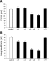
Summary data of adhesion force and integrated force with the FN-coated AFM probe in cardiomyocytes. The adhesion force (A) represents the first peak force, and the integrated force (B) represents the total area under the force-density distribution curves. Calculations are presented in materials and methods. Adhesion force was not changed in the presence of α3- or α5-integrin monoclonal antibodies alone, whereas the combination of α3- and α5-integrin monoclonal antibodies and β1-integrin monoclonal antibody decreased adhesion force. Integrated force, which provides a metric reflecting the average overall adhesiveness, was decreased by α3- or α5-integrin monoclonal antibodies and further decreased by the combination of α3- and α5-integrin monoclonal antibodies and β1-integrin monoclonal antibody (50 μM). Non-FN α11-integrin (60 nM) showed no significant effects on adhesion force and integrated force. *P < 0.05 vs. control (FN-coated AFM probe alone). n = 10 for each group.
Fig. 5.
Fig. 5.
Summary of results of adhesion characteristics of cardiomyocytes in the absence or presence of cytochalasin D or 2,3-butanedione monoxime (BDM). The adhesion force, probability, and cell stiffness were calculated as described in detail in materials and methods from the force curves obtained from cells under normal condition or cells incubated in the presence of cytochalasin D or BDM. Adhesion force (A) and adhesive probability (B) were not changed in the presence of cytochalasin D or BDM. Integrated force (C) and cell stiffness (D) were significantly decreased in the presence of cytochalasin D or BDM. *P < 0.05 vs. control. n = 10 for each group.
Fig. 6.
Fig. 6.
Summary of results of adhesion force in contracted (high-K+ solution), relaxed (low-Ca2+ solution), and high-K+ solution + BDM-treated cardiomyocytes. A–C: analyses of force-density plots of adhesion events during FN-coated probe retraction in cardiomyocytes. Adhesion to FN was enhanced during contraction or contraction with BDM compared with the relaxed state. The observed adhesion force and corresponding number of events in the experiments (50 curves/cell for a total of 500 curves) were plotted as histograms in contracted cells (A), relaxed cells (B), and high-K+ solution + BDM-treated cells (C). Solid lines represent the results that fitted with multiple Gaussian distributions. Insets: integrin-FN binding probabilities (solid bars) in contracted (A) and relaxed (B) cardiomyocytes and high-K+ solution + BDM treated cells (C). Top: AFM images of cardiomyocytes under each condition. n = 10 for each group.
Fig. 7.
Fig. 7.
Summary of results of cell adhesive and mechanical properties of cardiomyocytes in the contracted and relaxed states. The adhesion force (A), integrated force (B), and stiffness (C) of relaxed cells and high K+ solution + BDM-treated cells were significantly smaller than in contracted cells. In addition, the integrated force (B) and stiffness (C) of high-K+ solution + BDM-treated cells were significantly greater than in relaxed cells. *P < 0.05 vs. contracted cells; #P < 0.05 vs. relaxed cells. n = 10 for each group.
Fig. 8.
Fig. 8.
Simplified schematic representation of the FN-integrin-costamere axis. A: during relaxation, decreased adhesion force, probability of adhesion, and cell stiffness could be related to the decreased availability of activated integrin receptors within the costamere complex. B: in comparison, during contraction, increased adhesion force, probability of adhesion, and stiffness could be related to the increased availability of active integrin receptors and fortification of the costamere complex. The activation of integrins could hypothetically be related to inside-out ligand-activated signaling mechanisms (e.g., Ca2+) that can act to coordinate the contractile state with adhesion. Myofilaments: actin, myosin, tropomyosin, and troponin. ECM, extracellular matrix; +, activation; −, inhibition.

Similar articles

Cited by

References

    1. Ahumada GG, Saffitz JE. Fibronectin in rat heart: a link between cardiac myocytes and collagen. J Histochem Cytochem 32: 383–388, 1984 - PubMed
    1. Belke DD, Dillmann WH. Altered cardiac calcium handling in diabetes. Curr Hypertens Rep 6: 424–429, 2004 - PubMed
    1. Benoit M, Gabriel D, Gerisch G, Gaub HE. Discrete interactions in cell adhesion measured by single-molecule force spectroscopy. Nat Cell Biol 2: 313–317, 2000 - PubMed
    1. Brancaccio M, Hirsch E, Notte A, Selvetella G, Lembo G, Tarone G. Integrin signalling: the tug-of-war in heart hypertrophy. Cardiovasc Res 70: 422–433, 2006 - PubMed
    1. Brownlee M. The pathobiology of diabetic complications: a unifying mechanism. Diabetes 54: 1615–1625, 2005 - PubMed

Publication types

LinkOut - more resources