. 2010 Apr 1;66(Pt 4):379-85.
doi: 10.1107/S1744309110004318. Epub 2010 Mar 26.

X-ray structure of perdeuterated diisopropyl fluorophosphatase (DFPase): perdeuteration of proteins for neutron diffraction

Affiliations

X-ray structure of perdeuterated diisopropyl fluorophosphatase (DFPase): perdeuteration of proteins for neutron diffraction

Marc Michael Blum et al. Acta Crystallogr Sect F Struct Biol Cryst Commun. .

Abstract

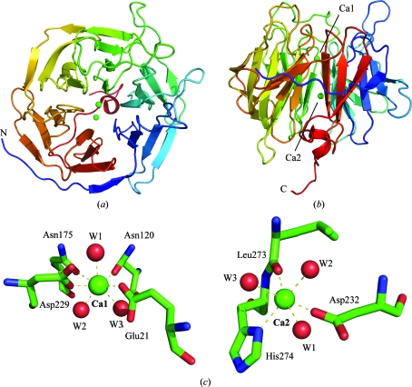
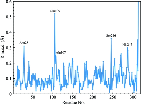
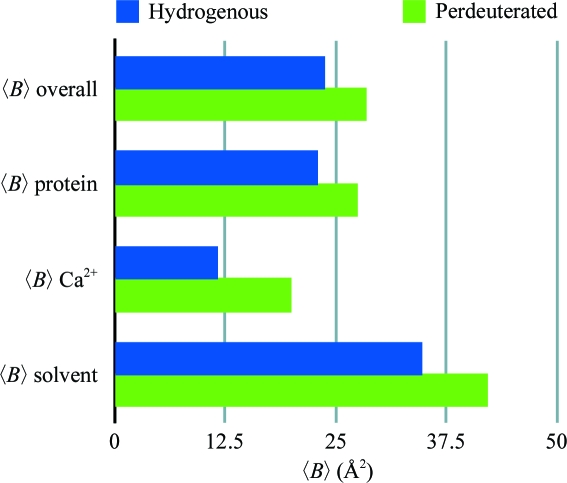
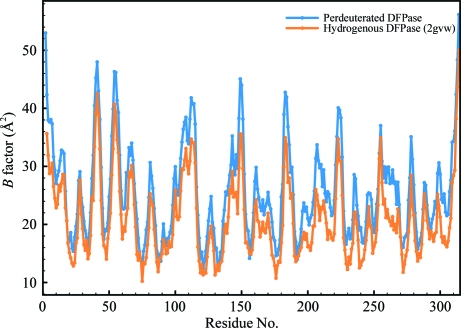
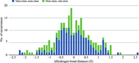
The signal-to-noise ratio is one of the limiting factors in neutron macromolecular crystallography. Protein perdeuteration, which replaces all H atoms with deuterium, is a method of improving the signal-to-noise ratio of neutron crystallography experiments by reducing the incoherent scattering of the hydrogen isotope. Detailed analyses of perdeuterated and hydrogenated structures are necessary in order to evaluate the utility of perdeuterated crystals for neutron diffraction studies. The room-temperature X-ray structure of perdeuterated diisopropyl fluorophosphatase (DFPase) is reported at 2.1 A resolution. Comparison with an independently refined hydrogenated room-temperature structure of DFPase revealed no major systematic differences, although the crystals of perdeuterated DFPase did not diffract neutrons. The lack of diffraction is examined with respect to data-collection and crystallographic parameters. The diffraction characteristics of successful neutron structure determinations are presented as a guideline for future neutron diffraction studies of macromolecules. X-ray diffraction to beyond 2.0 A resolution appears to be a strong predictor of successful neutron structures.


Figures

Figure 1
Figure 1
Overall structure of DFPase. (a) Top view along the propeller axis. (b) Side view showing the central structural calcium ion and the catalytic calcium ion at the bottom of the binding pocket. Colouring is according to primary sequence from blue (N-terminus) to red (C-terminus). (c) The coordination environment around the catalytic calcium (Ca1) and the structural calcium (Ca2).
Figure 2
Figure 2
Plot of Cα r.m.s.d. values between d-DFPase and h-DFPase.
Figure 3
Figure 3
Comparison of average B factors for d-DFPase and h-DFPase.
Figure 4
Figure 4
Plot of B factors for Cα atoms of individual residues in perdeuterated (blue) and hydrogenous (orange) DFPase.
Figure 5
Figure 5
Statistical distribution of hydrogen-bond distances shown as a histogram of the difference in distances between hydrogen-bonding pairs (perdeuterated distance − hydrogenated distance) for main-chain–main-chain and main-chain–side-chain interactions.
Figure 6
Figure 6
MALDI–TOF MS spectrum of hydrogenous and perdeuterated DFPase indicating full deuteration. The signals at 17 528 and 18 827 are protein molecules carrying a double charge.

References

    1. Adachi, M. et al. (2009). Proc. Natl Acad. Sci. USA, 106, 4641–4646. - PubMed
    1. Adams, P. D., Mustyakimov, M., Afonine, P. V. & Langan, P. (2009). Acta Cryst. D65, 567–573. - PMC - PubMed
    1. Arai, S., Chatake, T., Suzuki, N., Mizuno, H. & Niimura, N. (2004). Acta Cryst. D60, 1032–1039. - PubMed
    1. Artero, J.-B., Härtlein, M., McSweeney, S. & Timmins, P. (2005). Acta Cryst. D61, 1541–1549. - PubMed
    1. Bennett, B., Langan, P., Coates, L., Mustykimov, M., Schoenborn, B., Howell, E. E. & Dealwis, C. (2006). Proc. Natl Acad. Sci. USA, 103, 18493–18498. - PMC - PubMed

Publication types

Substances