. 2010 May;12(5):484-91.
doi: 10.1038/ncb2050. Epub 2010 Apr 11.

GEMC1 is a TopBP1-interacting protein required for chromosomal DNA replication

Affiliations

GEMC1 is a TopBP1-interacting protein required for chromosomal DNA replication

Alessia Balestrini et al. Nat Cell Biol. 2010 May.

Abstract

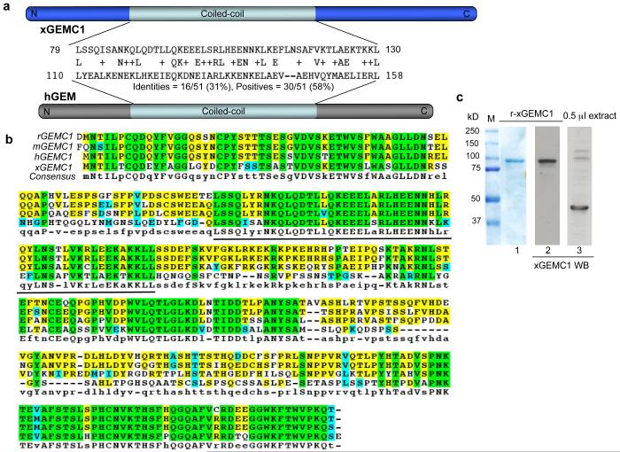
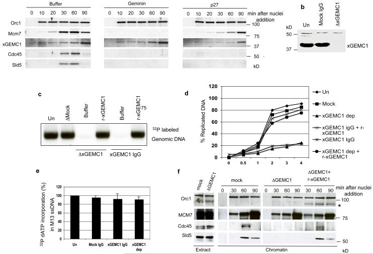
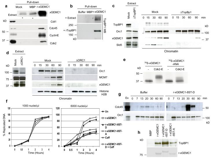
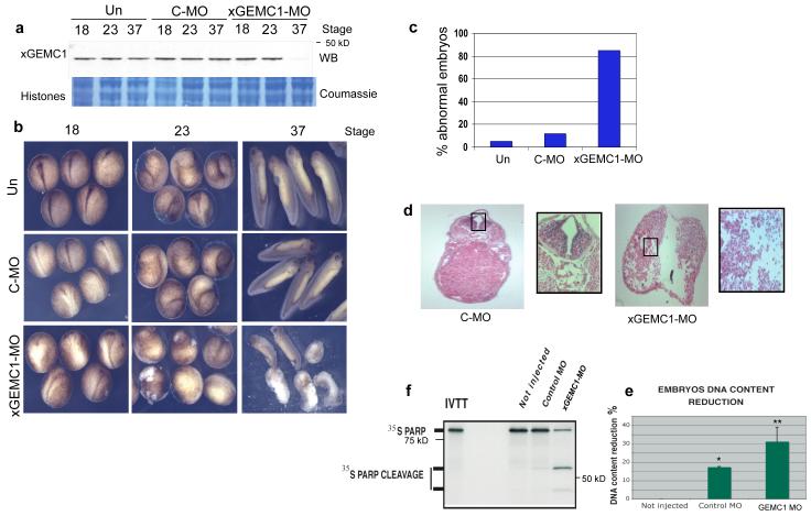
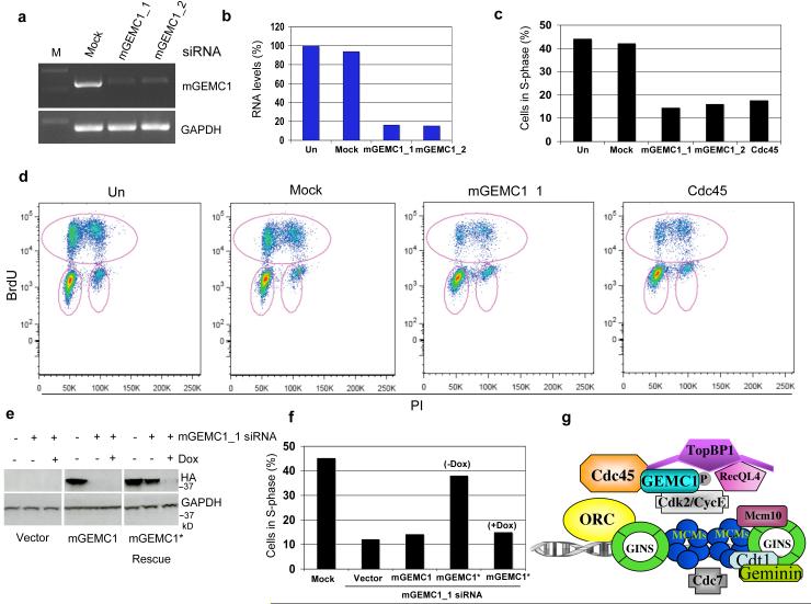
Many of the factors required for chromosomal DNA replication have been identified in unicellular eukaryotes. However, DNA replication is poorly understood in multicellular organisms. Here, we report the identification of GEMC1 (geminin coiled-coil containing protein 1), a novel vertebrate protein required for chromosomal DNA replication. GEMC1 is highly conserved in vertebrates and is preferentially expressed in proliferating cells. Using Xenopus laevis egg extract we show that Xenopus GEMC1 (xGEMC1) binds to the checkpoint and replication factor TopBP1, which promotes binding of xGEMC1 to chromatin during pre-replication complex (pre-RC) formation. We demonstrate that xGEMC1 interacts directly with replication factors such as Cdc45 and the kinase Cdk2-CyclinE, through which it is heavily phosphorylated. Phosphorylated xGEMC1 stimulates initiation of DNA replication, whereas depletion of xGEMC1 prevents the onset of DNA replication owing to the impairment of Cdc45 loading onto chromatin. Similarly, inhibition of GEMC1 expression with morpholino and siRNA oligos prevents DNA replication in embryonic and somatic vertebrate cells. These data suggest that GEMC1 promotes initiation of chromosomal DNA replication in multicellular organisms by mediating TopBP1- and Cdk2-dependent recruitment of Cdc45 onto replication origins.


Figures

Figure 1
Figure 1. Identification of GEMC1
a) Homology of the coiled-coil domain of xGEMC1 with human geminin (hGEM). b) Conservation of GEMC1 in vertebrates (R. Norvegicus rGEMC1 [XP_573306.2], M. Musculus mGEMC1 [NP_001013783], H. Sapiens hGEMC1 [NP_001140158.1] and X. laevis xGEMC1. Aminoacid identity in all species is in green. Line indicates coiled-coil domain. c) Expression of recombinant xGEMC1 (r-xGEMC1) fused to Maltose Binding Protein (MBP) monitored with coumassie stain (lane 1) or Western Blot (WB) (lane 2) using anti xGEMC1 antibodies. WB of endogenous xGEMC1 in egg extract is shown in lane 3. See Supplementary Fig 6 for uncropped gels shown in this figure.
Figure 2
Figure 2. Analysis of GEMC1 role in DNA replication
a) Chromatin binding of Orc1, MCM7, Cdc45, xGEMC1 and Sld5 at the indicated times after addition of sperm nuclei (3000 n/μl) to interphase egg extracts supplemented with buffer (Control), 80 nM geminin (GEM) or 1 μM p27 protein. The sample collected at 0 minutes was DNA free. b) WB of egg extract depleted with mock (ΔMock) or affinity purified anti xGEMC1 antibodies (ΔxGEMC1). c) DNA replication assay showing incorporation of α32P-dCTP in genomic DNA incubated in egg extract for 2 hours. From left to right sperm nuclei (1000 n-μl) were replicated in egg extracts that were untreated (Un), depleted with mock IgGs (Mock IgG, 80 ng/μl), depleted with affinity purified anti xGEMC1 IgGs (ΔxGEMC1) and supplemented with buffer (Buffer) or with 10 ng/μl xGEMC1 (r-xGEMC1) or incubated with affinity purified anti xGEMC1 IgGs (xGEMC1 IgG, 80 ng/μl) and supplemented with buffer (Buffer) or an excess of recombinant xGEMC1 (r-xGEMC1). DNA was isolated, run on agarose gel that was dried and exposed. d) Amount of replicated DNA measured by quantifying α32P-dATP incorporation over time using TCA precipitation (see Supplementary methods) in extracts treated as indicated in (c). The graph shows a typical result. e) Amount of α32P-dCTP incorporated in single stranded M13 phage DNA relative to untreated sample. Extracts were untreated (Un), supplemented with mock IgGs (Mock IgG, 80 ng/μl), with affinity purified anti xGEMC1 immunoglobulins (xGEMC1 IgG, 80 ng/μl) or xGEMC1 depleted (xGEMC1 dep). Experiment was repeated 3 times (n=3). Average results are shown. Error bars indicate SD. f) Chromatin binding of indicated proteins at different times after addition of sperm nuclei (3000 n/μl) to egg extracts that were mock (ΔMock) or xGEMC1 depleted in the absence (ΔxGEMC1) or in the presence (ΔxGEMC1+r-xGEMC1) of 10 ng/μl r-xGEMC1. (*) indicates binding of r-xGEMC1 to chromatin, which was probed with anti MBP antibodies. See Supplementary Fig 6 for uncropped gels shown in this figure.
Figure 3
Figure 3. Identification of GEMC1 interacting proteins
a) WB of indicated proteins on pull-downs performed with amylose resin that was untreated (Mock), pre-bound to MBP (MBP) or to xGEMC1 fused to MBP (r-xGEMC1) and subsequently incubated in egg extract. * indicates non-specific band. b) WB of TopBP1 on pull-downs performed with amylose resin that was untreated (Mock), pre-bound to MBP (MBP) or to GEMC1 fused to MBP (r-GEMC1) and incubated with extract (+ Extract) or with recombinant TopBP1. c) Chromatin binding of the indicated proteins at different times after addition of sperm nuclei (3000 n/μl) to egg extracts that were mock (Mock) or TopBP1 (ΔTopBP1) depleted. d) Chromatin binding of the indicated proteins at different times after addition of sperm nuclei (3000 n/μl) to interphase egg extracts that were mock (Mock) or Orc1 depleted (ΔORC1). e) Autoradiograph of 35S-labelled xGEMC1 (35S-GEMC1) or 35S-labelled xGEMC1 mutated in the Cyclin binding site (35S-xGEMC1-ANA) produced in reticulocyte lysates and incubated with buffer (−) or recombinant Cdk2-CyclinE complex (Cdk2-E). f) Amount of replicated DNA measured by quantifying α32P-dATP incorporation over time using TCA precipitation (see methods) at different times after the addition of 1000 or 6000 nuclei/μl to egg extracts supplemented with 300 ng/μl recombinant xGEMC1 (r-xGEMC1), 300 ng-μl xGEMC1-8ST-D (r-xGEMC1-8ST-D) carrying the serine and threonine residues phosphorylated by Cdk2 mutated to aspartate, 300 ng/μl xGEMC1-8ST-A protein (r-xGEMC1-8ST-A) with the same residues mutated to alanine, with 3 mM caffeine (Caff) or with 3 mM caffeine in the presence of 300 ng/μl xGEMC1-8ST-D protein (xGEMC1-8ST-D + Caff). The graphs show data from one representative experiment. xGEMC1 proteins used in these experiments were defective for the Cyclin-binding domain (R198NL to A198NA mutation) to avoid titration of endogenous Cdk2-Cyclin E complex. g) Cdc45 chromatin binding. Egg extracts were supplemented with buffer (Buffer) or 300 ng/μl xGEMC1-8ST-D mutant protein (r-xGEMC1-8ST-D). Orc1 was used as loading control. h) WB of TopBP1 (top panel) on pull-downs performed with amylose resin that was pre-bound to MBP (MBP), to MBP fused to wild type xGEMC1 (r-xGEMC1), to aspartate (r-xGEMC1-8ST-D) or to alanine substituted xGEMC1 (xGEMC1-8ST-A) (bottom panel, coumassie) and incubated with recombinant TopBP1. See Supplementary Fig 6 for uncropped gels shown in this figure.
Figure 4
Figure 4. Effects resulting from inhibition of GEMC1 expression in Xenopus embryos
a) WB of xGEMC1 extracted from Xenopus embryos that were untreated (Un) or injected with control (C-MO) and morpholino oligos complementary to xGEMC1 DNA sequence (xGEMC1-MO) taken at the indicated stages. Lower panel shows coumassie staining of histone proteins from total embryo lysates. b) Morphology of Xenopus embryos that were untreated (Un), injected with control (C-MO) or anti xGEMC1 morpholino oligos (xGEMC1-MO) taken at the indicated stages. c) Quantification of experiment shown in (b). 100 embryos were counted for each treatment. Graph shows a typical result. d) Sections of fixed Xenopus embryos injected with control (C-MO) or with anti xGEMC1 morpholino oligos (xGEMC1-MO) and stained with ematossilin-eosin. Area in the rectangle shows 5x magnification. e) DNA content reduction in embryos that were uninjected (Not injected), injected with control morpholino oligos (Control MO) or with anti xGEMC1 morpholino oligos (xGEMC1-MO). Data are mean ± SD of 3 independent experiments; * and ** P <0.001 compared with not injected control, t-test) f) Cleavage of 35S-labeled PARP induced by lysates of embryos that were uninjected (Not injected), injected with control morpholino oligos (Control MO) or anti xGEMC1 morpholino oligos (xGEMC1-MO). See Supplementary Fig 6 for un-cropped gels shown in this figure.
Figure 5
Figure 5. Effects resulting from inhibition of GEMC1 expression in mammalian cells
a) RT-PCR of mouse GEMC1 (mGEMC1) and GAPDH (GAPDH) mRNA derived from mouse NIH 3T3 fibroblasts that were treated with control (Mock) or mouse GEMC1 siRNA oligos (mGEMC1_1 and mGEMC1_2). b) Quantification of RNA levels shown in (a). The graph shows data from one representative experiment. c) Histograms relative to FACS analysis following incorporation of BrdU in mouse NIH3T3 cells that were untreated (Un), mock treated (Mock), transfected with two independent mouse GEMC1 siRNA (mGEMC1_1 or mGEMC1_2) or with Cdc45 siRNA oligos. The graph shows data from one representative experiment. d) Typical FACS profiles relative to experiments shown in (c). Circles in pink indicate BrdU incorporating cells. e) WB of MEF-3T3 cells stably transfected with empty TET-OFF vector (Vector), with TET-OFF vector expressing hemagglutinin epitope (HA) tagged mGEMC1 (mGEMC1) or with the same vector expressing siRNA resistant HA tagged mGEMC1 (mGEMC1* Rescue). Cells were subjected to silencing with mGEMC1_1 siRNA in the absence or presence of 1 μg/ml doxycycline (Dox), which represses the expression driven by the exogenous promoter present on the vector. After 96 hours cells were collected and processed for WB using anti HA and anti GAPDH antibodies as indicated. f) The histograms represent the percentage in S-phase of MEF-3T3 cells that were treated with mock (Mock) or mGEMC1 siRNA oligos (mGEMC1_1 siRNA). mGEMC1_1 siRNA treated cells were stably transfected with empty TET-OFF vector (Vector), with TET-OFF vector expressing HA tagged mGEMC1 (mGEMC1) or with the same vector expressing siRNA resistant HA tagged mGEMC1 (mGEMC1*) in the absence (−Dox) or in the presence (+Dox) of 1 μg/ml doxycycline. Cells were BrdUTP labelled and processed for FACS analysis. The graph shows data from one representative experiment. g) Model for GEMC1 function at replication origins (see text for explanation). See Supplementary Fig 6 for uncropped gels shown in this figure.

References

    1. Diffley JF. Regulation of early events in chromosome replication. Curr Biol. 2004;14:R778–786. - PubMed
    1. Takisawa H, Mimura S, Kubota Y. Eukaryotic DNA replication: from pre-replication complex to initiation complex. Curr Opin Cell Biol. 2000;12:690–696. - PubMed
    1. McGarry TJ, Kirschner MW. Geminin, an inhibitor of DNA replication, is degraded during mitosis. Cell. 1998;93:1043–1053. - PubMed
    1. Saxena S, et al. A dimerized coiled-coil domain and an adjoining part of geminin interact with two sites on Cdt1 for replication inhibition. Mol Cell. 2004;15:245–258. - PubMed
    1. Costanzo V, Gautier J. Xenopus cell-free extracts to study DNA damage checkpoints. Methods Mol Biol. 2004;241:255–267. - PubMed

Publication types

Associated data