Review
. 2010 May;17(5):530-8.
doi: 10.1038/nsmb.1779. Epub 2010 Apr 11.

Structures of influenza A proteins and insights into antiviral drug targets

Affiliations
Review

Structures of influenza A proteins and insights into antiviral drug targets

Kalyan Das et al. Nat Struct Mol Biol. 2010 May.

Abstract

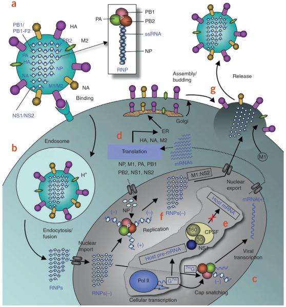
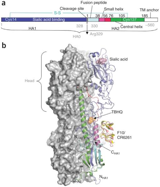
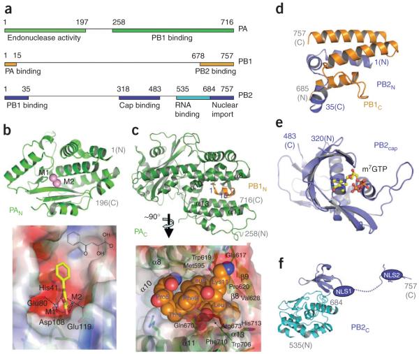
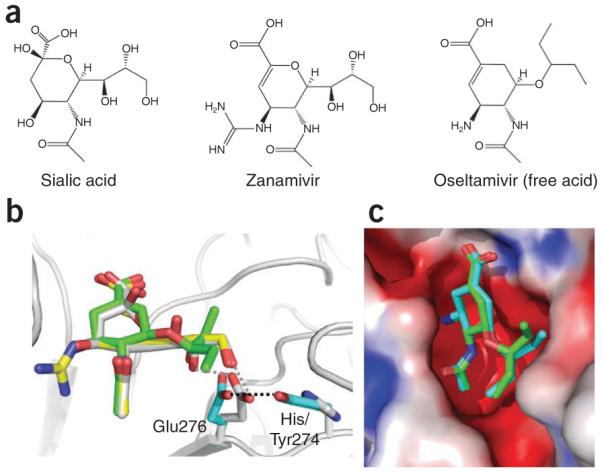
The world is currently undergoing a pandemic caused by an H1N1 influenza A virus, the so-called 'swine flu'. The H5N1 ('bird flu') influenza A viruses, now circulating in Asia, Africa and Europe, are extremely virulent in humans, although they have not so far acquired the ability to transfer efficiently from human to human. These health concerns have spurred considerable interest in understanding the molecular biology of influenza A viruses. Recent structural studies of influenza A virus proteins (or fragments) help enhance our understanding of the molecular mechanisms of the viral proteins and the effects of drug resistance to improve drug design. The structures of domains of the influenza RNA-dependent RNA polymerase and the nonstructural NS1A protein provide opportunities for targeting these proteins to inhibit viral replication.


Figures

Figure 1
Figure 1
Influenza A life cycle. (a) Influenza A virus has a lipid bilayer envelope, within which are eight RNA genomic segments, each of which is associated with the trimeric viral RNA polymerase (PB1, PB2, PA) and coated with multiple nucleoproteins (NPs) to form the vRNPs. The outer layer of the lipid envelope is spiked with multiple copies of HA, NA and a small number of M2, whereas the M1 molecules keep vRNPs attached to the inner layer. (b) The viral surface glycoprotein HA binds to the host cell-surface sialic acid receptors, and the virus is transported into the cell in an endocytic vesicle. The low pH in the endosome triggers a conformational change in the HA protein that leads to fusion of the viral and endosomal membranes. The low pH also triggers the flow of protons into the virus via the M2 ion channel, thereby dissociating the vRNPs from M1 matrix proteins. The vRNPs that are released into the cytoplasm are transported into the nucleus by recognition of the nuclear localization sequences (NLSs) on nucleoproteins only when the M1 molecules are dissociated. (c) In the nucleus, the viral polymerase initiates viral mRNA synthesis with 5′-capped RNA fragments cleaved from host pre-mRNAs. The PB2 subunit binds the 5′ cap of host pre-mRNAs, and the endonuclease domain in PA subunit cleaves the pre-mRNA 10–13 nucleotides downstream from the cap. Viral mRNA transcription is subsequently initiated from the cleaved 3′ end of the capped RNA segment,. This ‘cap snatching’ occurs on nascent pre-mRNAs. (d) Viral mRNAs are transported to the cytoplasm for translation into viral proteins. The surface proteins HA, M2 and NA are processed in the endoplasmic reticulum (ER), glycosylated in the Golgi apparatus and transported to the cell membrane. (e) The NS1 protein of influenza A virus serves a critical role in suppressing the production of host mRNAs by inhibiting the 3′-end processing of host pre-mRNAs,, consequently blocking the production of host mRNAs, including interferon-β mRNAs. Unlike host pre-mRNAs, the viral mRNAs do not require 3′-end processing by the host cell machinery. Therefore, the viral mRNAs are transported to the cytoplasm, whereas the host mRNA synthesis is predominantly blocked. (f) The viral polymerase is responsible for not only capped RNA-primed mRNA synthesis but also unprimed replication of vRNAs in steps (−) vRNA → (+) cRNA → (−) vRNA. The nucleoprotein molecules are required for these two steps of replication and are deposited on the cRNA and vRNA during RNA synthesis. The resulting vRNPs are subsequently transported to the cytoplasm, mediated by a M1–NS2 complex that is bound to the vRNPs; NS2 interacts with human CRM1 protein that exports the vRNPs from the nucleus. (g) The vRNPs reach the cell membrane to be incorporated into new viruses (reviewed in ref. 89) that are budded out. The HA and NA proteins in new viruses contain terminal sialic acids that would cause the viruses to clump together and adhere to the cell surface. The NA of newly formed viruses cleaves these sialic acid residues, thereby releasing the virus from the host cell.
Figure 2
Figure 2
Structural arrangement of HA trimers at prefusion state. (a) The HA precursor (HA0) is cleaved into sialic acid receptor binding domain (HA1) and ectodomain (HA2) that remain disulfide linked; TM, transmembrane anchor. (b) HA (HA1 + HA2) exists in trimeric form with a combined mass of ~220 kDa. Two HA molecules in a trimer are represented by their combined molecular surface, whereas the third one is shown in a ribbon representation, color coded according to panel a. The head of the HA1 chain (blue), which binds sialic acid receptors, has essentially all the antigenic sites against which human antibodies are directed. The HA1 head region is closely associated with the central coiled-coil trimeric α-helices (green). At fusion (low) pH, all three HA1 heads swing away from HA2 (ref. 91). The loops (red) connecting the coiled-coil stem to the fusion peptides are refolded to form helical structures that extend the coiled coil and expose the fusion peptides at the N-terminal end of HA2 (NHA2), which then contacts the endosome membrane for the fusion step. Binding of the small-molecule inhibitor TBHQ (orange) at the HA:HA interface or binding of human monoclonal antibody CR6261 (yellow) or F10 (salmon) to a conserved region adjacent to the TBHQ pocket inhibits the low pH conformational change of HA.
Figure 3
Figure 3
Structure, function and inhibition of the proton channel M2 protein of influenza A. (a) The vRNPs are attached to the lipid bilayer membrane via M1 matrix proteins. Influx of the protons from endosome to virus through M2 channels releases vRNPs. (b) The adamantanes (amantadine and rimantadine) inhibit the proton flow through the tetrameric M2 channel. (c) X-ray and NMR structures of M2 channel. (i) crystal structure of the transmembrane (TM) domain of M2–amantadine complex at pH 5.3 in which the drug (orange) binds M2 near Ser31 (ref. 17); (ii) solution NMR structure of M2–rimantadine complex at pH 7.5 reveals that the drug binds the individual M2 TM helix near Trp41 (ref. 18) (this structure also contains the C terminus cytoplasmic tail helix, not shown in the figure); (iii) solid-state NMR structure of M2–amantadine complex at pH 7.5 (ref. 19) reveals the drug binding to the proton channel, which is analogous to that in structure i (however, the spatial arrangements of the TM helices are different); and (iv) the arrangement of M2 TM helices as revealed by a solid-state NMR study of amantadine-bound M2 TM helix at pH 8.8 (ref. 92). The side chains of Ser31 (gray), His37 (cyan) and Trp41 (yellow) are shown in each structure.
Figure 4
Figure 4
Structure of nucleoprotein. A nucleoprotein monomer has a crescent-shaped body and a tail loop. Comparison of two available crystal structures of nucleoprotein, (PDB 2IQH and 2Q06) indicates the repositioning of the tail loop; the domain names are swapped to represent the current orientation. The tail loop of the neighboring nucleoprotein molecule (green) binds to a cavity at the back of the body domain. A zoomed view showing the binding of the tip of the tail loop (residues 408–419) to the loop-binding cavity, a potential site for drug design; the residues of the loop are labeled.
Figure 5
Figure 5
Structurally characterized fragments of influenza A polymerase complex (P-complex) that is involved in transcription and replication. (a) The heterotrimetric polymerase has subunits PA, PB1 and PB2 with a combined mass of ~250 kDa. The segments that have structural information are represented as colored bars—the colors correspond to the respective structures in the surrounding panels. (b) Ribbon representation of PAN domain (above) and electrostatic potential surface (below) of the endonuclease active site region,. We have docked an influenza endonuclease inhibitor, 2,4-dioxo-4-phenylbutanoic acid (yellow), that coordinates with the active-site metal ions (M1 and M2). The large cavity to which the phenyl ring points is a potential target for designing inhibitors with high binding affinity. (c) Ribbon representation (above) of PAC domain (green) in complex with an N-terminal helix of PB1N (gold) and the molecular surface of a section of the structure, (below). The PB1N helix (P5TLLFLK11), shown in space-filling model, occupies a pocket which is a likely target for small-molecule inhibitors of PAC:PB1N dimerization. The PB1N helix has both hydrophobic and hydrogen-bond interactions with the PAC pocket residues that can be exploited for the inhibitor design. (d) Structure of PB1C–PB2N complex shows a ‘revolver-shaped’ helix bundle. (e) The structure of the CAP (m7GTP) binding moiety PB2cap in complex with a m7GTP molecule. (f) C-terminal region of PB2 contains a bipartite nuclear localized sequence (NLS) domain (blue), the structure of which was determined in complex with importin α5 (ref. 48). The adjacent domain (cyan) contains an RNA-binding cleft.
Figure 6
Figure 6
Structures of N- and C-terminal domains of influenza A nonstructural protein NS1A and complexes. (a) The N-terminal domain (NS1AN) forms a highly stable dimer that binds dsRNA in a non–sequence specific manner. (b) The C-terminal effector domain (NS1AC) has been shown to bind F2F3 zinc fingers of human CPSF30 (ref. 62); this function of NS1A is essential to inhibit 3′-end processing of cellular pre-mRNAs. (c) The dimer interface in apo NS1AC. (d) Upon binding of CPSF30, the NS1AC domain undergoes conformational changes (from c to d) to create a hydrophobic pocket that accommodates three aromatic residues of the F3 zinc finger. The pocket region is highly conserved in influenza A viruses and is a promising target for anti–influenza A drugs that would block a key host and viral protein interaction. In apo structures,, as represented in c, the pocket does not exist, and a hydrophobic patch at the region is hidden at an intermolecular interface, primarily due to the conserved Trp187 side chain.
Figure 7
Figure 7
Binding of sialic acid–mimic drugs zanamivir and oseltamivir to neuraminidase (NA). (a,b) Chemical structures (a) and superposition of NA structures (b) show highly similar modes of binding for zanamivir (yellow) and oseltamivir (green) to sialic acid (gray) substrate, which reflects the influence of structures in the discovery of these drugs,. Recent crystal structures of oseltamivir-resistant mutant NA complexes show how the virus uses the NA H274Y mutation to reposition Glu276, which discriminates the l-ethylpropoxy group of oseltamivir from the glycerol moiety of the substrate. The repositioned Glu276 side chain would develop steric conflict with the l-ethylpropoxy group of oseltamivir, whereas it could still maintain favorable interactions with the glycerol part, common to both sialic acid and zanamivir. (c) Comparison of the modes of binding of oseltamivir to wild-type and H267Y mutant NA (shown in green and cyan, respectively). The H264Y mutation affects the positioning of the l-ethylpropoxy group of oseltamivir. The rearrangement of l-ethylpropoxy group of oseltamivir is associated with loss of inhibitor-protein interactions, resulting in a significant drug resistance.

Similar articles

Cited by

References

    1. Reid AH, Taubenberger JK, Fanning TG. The 1918 Spanish influenza: integrating history and biology. Microbes Infect. 2001;3:81–87. - PubMed
    1. Chowell G, et al. Severe respiratory disease concurrent with the circulation of H1N1 influenza. N. Engl. J. Med. 2009;361:674–679. - PubMed
    1. Garten RJ, et al. Antigenic and genetic characteristics of swine-origin 2009 A(H1N1) influenza viruses circulating in humans. Science. 2009;325:197–201. - PMC - PubMed
    1. Lamb RA, Krug RM. Orthomyxoviridae: the viruses and their replication. In: Knipe DM, Howley PM, editors. Fields Virology. 4th edn. Lippincott, Williams & Wilkins; Philadelphia, USA: 2001. pp. 1487–1532.
    1. Wright PF, Webster RG. Orthomyxoviruses. In: Knipe DM, Howley PM, editors. Fields Virology. 4th edn. Lippincott, Williams & Wilkins; Philadelphia, USA: 2001. pp. 1533–1579.

Publication types