

Simultaneous dual-radionuclide myocardial perfusion imaging with a solid-state dedicated cardiac camera
- PMID: 20383705
- PMCID: PMC3108881
- DOI: 10.1007/s00259-010-1441-1
Simultaneous dual-radionuclide myocardial perfusion imaging with a solid-state dedicated cardiac camera
Abstract
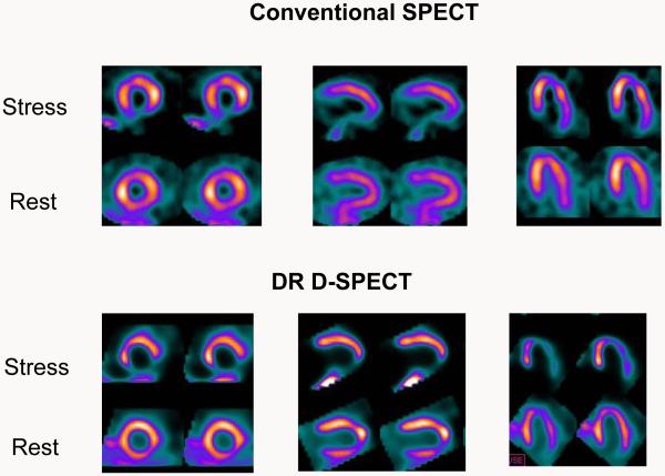
Purpose: We compared simultaneous dual-radionuclide (DR) stress and rest myocardial perfusion imaging (MPI) with a novel solid-state cardiac camera and a conventional SPECT camera with separate stress and rest acquisitions.
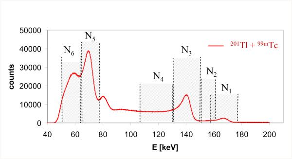
Methods: Of 27 consecutive patients recruited, 24 (64.5+/-11.8 years of age, 16 men) were injected with 74 MBq of (201)Tl (rest) and 250 MBq (99m)Tc-MIBI (stress). Conventional MPI acquisition times for stress and rest are 21 min and 16 min, respectively. Rest (201)Tl for 6 min and simultaneous DR 15-min list mode gated scans were performed on a D-SPECT cardiac scanner. In 11 patients DR D-SPECT was performed first and in 13 patients conventional stress (99m)Tc-MIBI SPECT imaging was performed followed by DR D-SPECT. The DR D-SPECT data were processed using a spill-over and scatter correction method. DR D-SPECT images were compared with rest (201)Tl D-SPECT and with conventional SPECT images by visual analysis employing the 17-segment model and a five-point scale (0 normal, 4 absent) to calculate the summed stress and rest scores. Image quality was assessed on a four-point scale (1 poor, 4 very good) and gut activity was assessed on a four-point scale (0 none, 3 high).
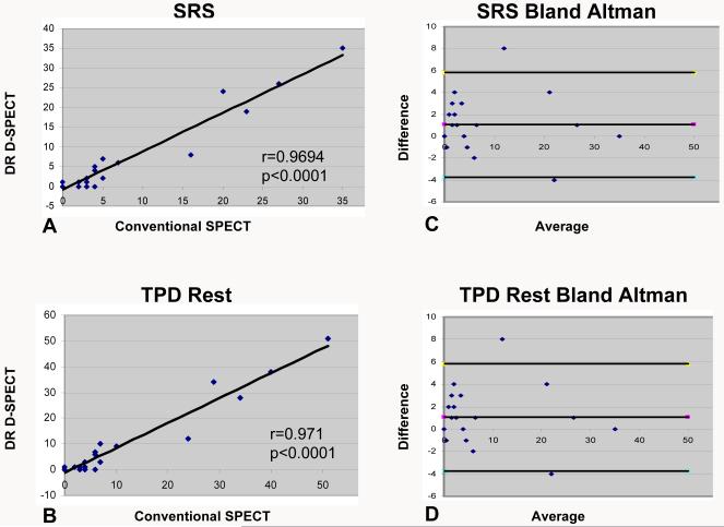
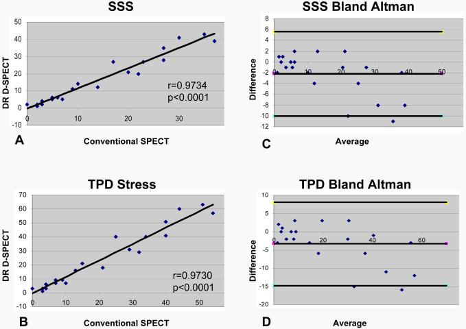
Results: Conventional MPI studies were abnormal at stress in 17 patients and at rest in 9 patients. In the 17 abnormal stress studies DR D-SPECT MPI showed 113 abnormal segments and conventional MPI showed 93 abnormal segments. In the nine abnormal rest studies DR D-SPECT showed 45 abnormal segments and conventional MPI showed 48 abnormal segments. The summed stress and rest scores on conventional SPECT and DR D-SPECT were highly correlated (r=0.9790 and 0.9694, respectively). The summed scores of rest (201)Tl D-SPECT and DR-DSPECT were also highly correlated (r=0.9968, p<0.0001 for all). In six patients stress perfusion defects were significantly larger on stress DR D-SPECT images, and five of these patients were imaged earlier by D-SPECT than by conventional SPECT.
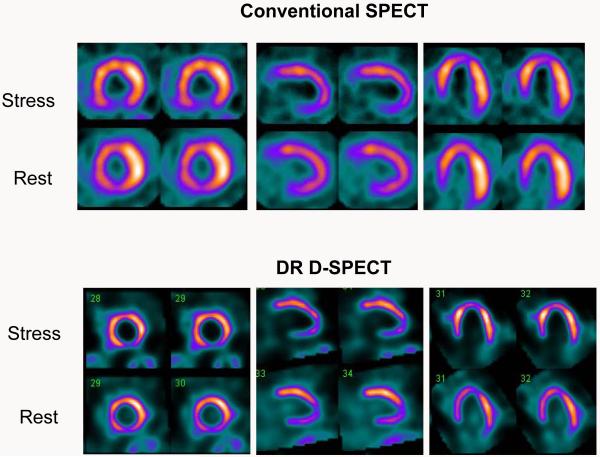
Conclusion: Fast and high-quality simultaneous DR MPI is feasible with D-SPECT in a single imaging session with comparable diagnostic performance and image quality to conventional SPECT and to a separate rest (201)Tl D-SPECT acquisition.
Figures
Comment in
-
Dedicated cardiac cameras: a new option for nuclear myocardial perfusion imaging.Eur J Nucl Med Mol Imaging. 2010 Aug;37(9):1706-9. doi: 10.1007/s00259-010-1526-x. Eur J Nucl Med Mol Imaging. 2010. PMID: 20593283 No abstract available.
References
-
- Patton JA, Slomka P, Germano G, Berman D. Recent technological advances in nuclear cardiology. J Nucl Cardiol. 2007;14:501–513. - PubMed
-
- Slomka PJ, Patton JA, Berman DS, Germano G. Advances in technical aspects of myocardial perfusion SPECT imaging. J Nucl Cardiol. 2009;16:255–276. - PubMed
-
- Gambhir SS, Berman DS, Ziffer J, Nagler M, Sandler M, Patton J, Hutton B, Sharir T, Ben-Haim S, Ben-Haim S. A novel high-sensitivity rapid-acquisition single-photon cardiac imaging camera. J Nucl Med. 2009;50:635–643. - PubMed
-
- Erlandsson K, Kacperski K, Van Gramberg D, Hutton BF. Performance evaluation of D-SPECT: a novel SPECT system for nuclear cardiology. Phys Med Biol. 2009;54:2635–2649. - PubMed
-
- Sharir T, Ben-Haim S, Merzon K, Prochorov V, Dickman D, Ben-Haim S, Berman DS. High-speed myocardial perfusion imaging: Initial clinical comparison with conventional dual detector Anger camera imaging. J Am Coll Cardiol Img. 2008;1:156–163. - PubMed
Publication types
MeSH terms
Grants and funding
LinkOut - more resources
Full Text Sources
