

A technique optimization protocol and the potential for dose reduction in digital mammography
- PMID: 20384232
- PMCID: PMC2830259
- DOI: 10.1118/1.3276732
A technique optimization protocol and the potential for dose reduction in digital mammography
Abstract
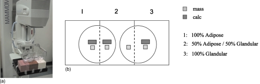
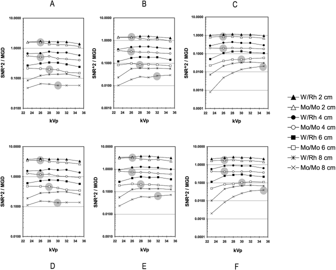
Digital mammography requires revisiting techniques that have been optimized for prior screen/film mammography systems. The objective of the study was to determine optimized radiographic technique for a digital mammography system and demonstrate the potential for dose reduction in comparison to the clinically established techniques based on screen- film. An objective figure of merit (FOM) was employed to evaluate a direct-conversion amorphous selenium (a-Se) FFDM system (Siemens Mammomat Novation(DR), Siemens AG Medical Solutions, Erlangen, Germany) and was derived from the quotient of the squared signal-difference-to-noise ratio to mean glandular dose, for various combinations of technique factors and breast phantom configurations including kilovoltage settings (23-35 kVp), target/filter combinations (Mo-Mo and W-Rh), breast-equivalent plastic in various thicknesses (2-8 cm) and densities (100% adipose, 50% adipose/50% glandular, and 100% glandular), and simulated mass and calcification lesions. When using a W-Rh spectrum, the optimized FOM results for the simulated mass and calcification lesions showed highly consistent trends with kVp for each combination of breast density and thickness. The optimized kVp ranged from 26 kVp for 2 cm 100% adipose breasts to 30 kVp for 8 cm 100% glandular breasts. The use of the optimized W-Rh technique compared to standard Mo-Mo techniques provided dose savings ranging from 9% for 2 cm thick, 100% adipose breasts, to 63% for 6 cm thick, 100% glandular breasts, and for breasts with a 50% adipose/50% glandular composition, from 12% for 2 cm thick breasts up to 57% for 8 cm thick breasts.
Figures
References
-
- Muller S., “Full-field digital mammography designed as a complete system,” Eur. Radiol. ZZZZZZ 31, 25–34 (1997). - PubMed
Publication types
MeSH terms
LinkOut - more resources
Full Text Sources
Other Literature Sources
Medical
