Long-term oncological outcomes of ovarian serous carcinomas with psammoma bodies: a novel insight into the molecular pathogenesis of ovarian epithelial carcinoma
- PMID: 20384630
- PMCID: PMC11158184
- DOI: 10.1111/j.1349-7006.2010.01556.x
Long-term oncological outcomes of ovarian serous carcinomas with psammoma bodies: a novel insight into the molecular pathogenesis of ovarian epithelial carcinoma
Abstract
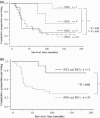
A two-tier system in which ovarian epithelial carcinomas are subdivided into type I and type II tumors has been proposed on the basis of recent molecular pathogenesis findings. Type I tumors, unrelated to tumor protein p53 (TP53) mutations, show favorable prognosis in a slow step-wise process, whereas type II tumors, related to TP53 mutations, contribute to poor prognosis. Ovarian serous carcinomas with excessive psammoma bodies behave like type I tumors. However, their etiology and prognostic significance remain obscure. The objective of the present study was to evaluate the characteristic features and potential relevance of psammoma bodies to the clinical outcome of 44 patients with serous carcinomas with long-term follow-up. The 5- and 10-year survival rates were significantly different between the serous carcinomas with less than 5% area of psammoma bodies and those at least 5% area (P < 0.01). All tumors with at least 5% area were both diploid and immunohistochemically negative for TP53 mutations. All patients with these tumors, including eight with International Federation of Gynecology and Obstetrics (FIGO) stages III or IV disease, survived more than 5 years and their 10-year survival rate was 76%. In multivariate analysis using clinical parameters, the apparent existence of psammoma bodies was an indication to view serous carcinomas as type I tumors with long-term survival. Our results suggested that the formation of psammoma bodies is associated with increased apoptotic tumor cell death related to normal TP53 function. The pathological findings of psammoma bodies might contribute to the consideration of pathogenesis and to the development of prognostic prediction rules for serous carcinomas.
Figures


Similar articles
-
[Clinicopathologic analysis and expression of cyclin D1 and p53 of ovarian borderline tumors and carcinomas].Zhonghua Fu Chan Ke Za Zhi. 2007 Apr;42(4):227-32. Zhonghua Fu Chan Ke Za Zhi. 2007. PMID: 17631760 Chinese.
-
Clear cell carcinomas of the ovary have poorer outcomes compared with serous carcinomas: Results from a single-center Taiwanese study.J Formos Med Assoc. 2018 Feb;117(2):117-125. doi: 10.1016/j.jfma.2017.03.007. Epub 2017 Apr 4. J Formos Med Assoc. 2018. PMID: 28389144
-
TP53 mutations are common in all subtypes of epithelial ovarian cancer and occur concomitantly with KRAS mutations in the mucinous type.Exp Mol Pathol. 2013 Oct;95(2):235-41. doi: 10.1016/j.yexmp.2013.08.004. Epub 2013 Aug 18. Exp Mol Pathol. 2013. PMID: 23965232
-
Low-grade serous ovarian cancer: A review.Gynecol Oncol. 2016 Nov;143(2):433-438. doi: 10.1016/j.ygyno.2016.08.320. Epub 2016 Aug 28. Gynecol Oncol. 2016. PMID: 27581327 Review.
-
Extrauterine Pelvic Serous Carcinomas: Current Update on Pathology and Cross-sectional Imaging Findings.Radiographics. 2016 May-Jun;36(3):918-32. doi: 10.1148/rg.2016150130. Radiographics. 2016. PMID: 27163599 Review.
Cited by
-
A binary histologic grading system for ovarian serous carcinoma is an independent prognostic factor: a population-based study of 4317 women diagnosed in Denmark 1978-2006.Gynecol Oncol. 2012 Jun;125(3):655-60. doi: 10.1016/j.ygyno.2012.02.028. Epub 2012 Feb 24. Gynecol Oncol. 2012. PMID: 22370600 Free PMC article.
-
An evolving story of the metastatic voyage of ovarian cancer cells: cellular and molecular orchestration of the adipose-rich metastatic microenvironment.Oncogene. 2019 Apr;38(16):2885-2898. doi: 10.1038/s41388-018-0637-x. Epub 2018 Dec 19. Oncogene. 2019. PMID: 30568223 Free PMC article. Review.
-
Low Grade Ovarian Serous Carcinoma - A Clinical-Morphologic Study.Curr Health Sci J. 2019 Jan-Mar;45(1):42-46. doi: 10.12865/CHSJ.45.01.05. Epub 2019 Mar 31. Curr Health Sci J. 2019. PMID: 31297261 Free PMC article.
-
Ovarian Cancer Stemness: Biological and Clinical Implications for Metastasis and Chemotherapy Resistance.Cancers (Basel). 2019 Jun 28;11(7):907. doi: 10.3390/cancers11070907. Cancers (Basel). 2019. PMID: 31261739 Free PMC article. Review.
-
CD44 variant 6 is correlated with peritoneal dissemination and poor prognosis in patients with advanced epithelial ovarian cancer.Cancer Sci. 2015 Oct;106(10):1421-8. doi: 10.1111/cas.12765. Epub 2015 Sep 21. Cancer Sci. 2015. PMID: 26250934 Free PMC article.
References
-
- Jemal A, Siegel R, Ward E, Murray T, Xu J, Thun MJ. Cancer statistics, 2007. CA Cancer J Clin 2007; 57: 43–66. - PubMed
-
- Sorbe B, Frankendal B, Veress B. Importance of histologic grading in the prognosis of epithelial ovarian carcinoma. Obstet Gynecol 1982; 59: 576–82. - PubMed
-
- Sigurdsson K, Alm P, Gullberg B. Prognostic factors in malignant epithelial ovarian tumors. Gynecol Oncol 1983; 15: 370–80. - PubMed
-
- Large JM, Weinberg DS, Huettner PC, Mark SD. Flow cytometric analysis of nuclear DNA content in ovarian tumors: Association of ploidy with tumor type, histologic grade, and clinical stage. Cancer 1992; 69: 2668–75. - PubMed
-
- Kim YT, Zhao M, Kim SH, Lee CS, Kim JH, Kim JW. Prognostic significance of DNA quantification by flow cytometry in ovarian tumors. Int J Gynaecol Obstet 2005; 88: 286–91. - PubMed
Publication types
MeSH terms
LinkOut - more resources
Full Text Sources
Medical
Research Materials
Miscellaneous

