

Brain development in children with new onset epilepsy: a prospective controlled cohort investigation
- PMID: 20384719
- PMCID: PMC4120892
- DOI: 10.1111/j.1528-1167.2010.02563.x
Brain development in children with new onset epilepsy: a prospective controlled cohort investigation
Abstract
Purpose: To characterize prospective neurodevelopmental changes in brain structure in children with new and recent-onset epilepsy compared to healthy controls.
Methods: Thirty-four healthy controls (mean age 12.9 years) and 38 children with new/recent-onset idiopathic epilepsy (mean age 12.9 years) underwent 1.5 T magnetic resonance imaging (MRI) at baseline and 2 years later. Prospective changes in total cerebral and lobar gray and white matter volumes were compared within and between groups.
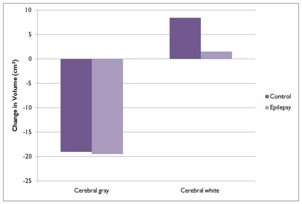
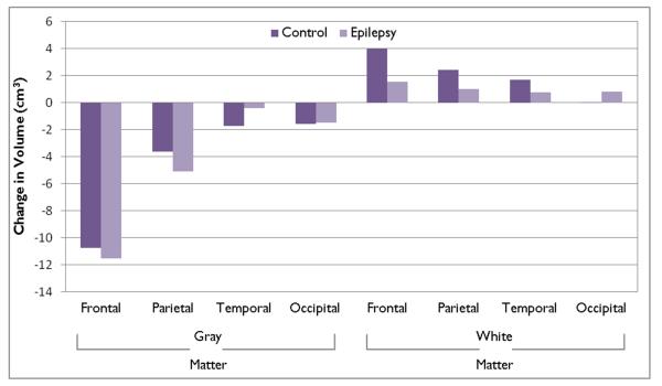
Results: Prospective changes in gray matter volume were comparable for the epilepsy and control groups, with significant (p < 0.0001) reduction in total cerebral gray matter, due primarily to significant (p < 0.001) reductions in frontal and parietal gray matter. Prospective white matter volume changes differed between groups. Controls exhibited a significant (p = 0.0012) increase in total cerebral white matter volume due to significant (p < 0.001) volume increases in the frontal, parietal, and temporal lobes. In contrast, the epilepsy group exhibited nonsignificant white matter volume change in the total cerebrum (p = 0.51) as well as across all lobes (all p's > 0.06). The group by white matter volume change interactions were significant for total cerebrum (p = 0.04) and frontal lobe (p = 0.04).
Discussion: Children with new and recent-onset epilepsy exhibit an altered pattern of brain development characterized by delayed age-appropriate increase in white matter volume. These findings may affect cognitive development through reduced brain connectivity and may also be related to the impairments in executive function commonly reported in this population.
Wiley Periodicals, Inc. © 2010 International League Against Epilepsy.
Figures
Similar articles
-
Children with new-onset epilepsy: neuropsychological status and brain structure.Brain. 2006 Oct;129(Pt 10):2609-19. doi: 10.1093/brain/awl196. Epub 2006 Aug 23. Brain. 2006. PMID: 16928696
-
Brain structure and aging in chronic temporal lobe epilepsy.Epilepsia. 2012 Jun;53(6):1033-43. doi: 10.1111/j.1528-1167.2012.03447.x. Epub 2012 Apr 3. Epilepsia. 2012. PMID: 22471353 Free PMC article.
-
[Reduction of gray and white matters in patients with temporal lobe epilepsy and its correlation with disease duration].Zhongguo Yi Xue Ke Xue Yuan Xue Bao. 2013 Jun;35(3):286-93. doi: 10.3881/j.issn.1000-503X.2013.03.009. Zhongguo Yi Xue Ke Xue Yuan Xue Bao. 2013. PMID: 23827066 Chinese.
-
Are there progressive brain changes in schizophrenia? A meta-analysis of structural magnetic resonance imaging studies.Biol Psychiatry. 2011 Jul 1;70(1):88-96. doi: 10.1016/j.biopsych.2011.01.032. Epub 2011 Mar 31. Biol Psychiatry. 2011. PMID: 21457946
-
Neurodevelopmental origins of self-limiting rolandic epilepsy: Systematic review of MR imaging studies.Epilepsia Open. 2021 Jun;6(2):310-322. doi: 10.1002/epi4.12468. Epub 2021 Mar 2. Epilepsia Open. 2021. PMID: 34033258 Free PMC article.
Cited by
-
Functional and Structural Network Disorganizations in Typical Epilepsy With Centro-Temporal Spikes and Impact on Cognitive Neurodevelopment.Front Neurol. 2019 Aug 29;10:809. doi: 10.3389/fneur.2019.00809. eCollection 2019. Front Neurol. 2019. PMID: 31555191 Free PMC article. Review.
-
The relationship between neuromagnetic networks and cognitive impairment in self-limited epilepsy with centrotemporal spikes.Epilepsia Open. 2025 Jun;10(3):842-854. doi: 10.1002/epi4.70044. Epub 2025 Apr 15. Epilepsia Open. 2025. PMID: 40231835 Free PMC article.
-
Evaluation of cortical thickness and brain volume on 3 Tesla magnetic resonance imaging in children with frontal lobe epilepsy.Neurol Sci. 2020 Apr;41(4):825-833. doi: 10.1007/s10072-019-04135-4. Epub 2019 Dec 4. Neurol Sci. 2020. PMID: 31802343
-
Evaluation of subcortical grey matter abnormalities in patients with MRI-negative cortical epilepsy determined through structural and tensor magnetic resonance imaging.BMC Neurol. 2014 May 14;14:104. doi: 10.1186/1471-2377-14-104. BMC Neurol. 2014. PMID: 24885823 Free PMC article.
-
Stress phenotypes in epilepsy: impact on cognitive functioning and quality of life.Front Psychol. 2023 Jun 14;14:1100101. doi: 10.3389/fpsyg.2023.1100101. eCollection 2023. Front Psychol. 2023. PMID: 37388654 Free PMC article.
References
-
- World Medical Association World Medical Association Declaration of Helsinki. The Journal of Law, Medicine & Ethics. 1991;19:264–265. - PubMed
-
- Alexander AL, Lee JE, Lazar M, Boudos R, DuBray MB, Oakes TR, Miller JN, Lu J, Jeong EK, McMahon WM, Bigler ED, Lainhart JE. Diffusion tensor imaging of the corpus callosum in Autism. Neuroimage. 2007;34:61–73. - PubMed
-
- Andreasen NC, Rajarethinam R, Cizadlo T, Arndt S, Swayze VW, 2nd, Flashman LA, O’Leary DS, Ehrhardt JC, Yuh WT. Automatic atlas-based volume estimation of human brain regions from MR images. J Comput Assist Tomogr. 1996;20:98–106. - PubMed
-
- Betting LE, Mory SB, Li LM, Lopes-Cendes I, Guerreiro MM, Guerreiro CA, Cendes F. Voxel-based morphometry in patients with idiopathic generalized epilepsies. Neuroimage. 2006a;32:498–502. - PubMed
-
- Betting LE, Mory SB, Lopes-Cendes I, Li LM, Guerreiro MM, Guerreiro CA, Cendes F. MRI reveals structural abnormalities in patients with idiopathic generalized epilepsy. Neurology. 2006b;67:848–852. - PubMed
Publication types
MeSH terms
Grants and funding
LinkOut - more resources
Full Text Sources
Other Literature Sources
Medical
