

Lack of oxygen in articular cartilage: consequences for chondrocyte biology
- PMID: 20384821
- PMCID: PMC2965894
- DOI: 10.1111/j.1365-2613.2010.00707.x
Lack of oxygen in articular cartilage: consequences for chondrocyte biology
Erratum in
- Int J Exp Pathol. 2010 Jun;91(3):302
Abstract
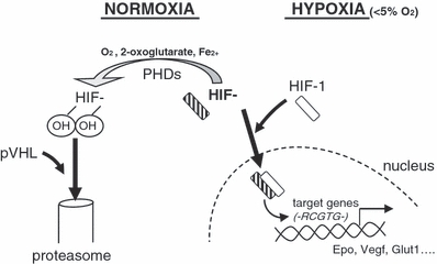
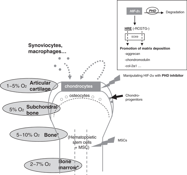
Controlling the chondrocytes phenotype remains a major issue for cartilage repair strategies. These cells are crucial for the biomechanical properties and cartilage integrity because they are responsible of the secretion of a specific matrix. But chondrocyte dedifferentiation is frequently observed in cartilage pathology as well as in tissue culture, making their study more difficult. Given that normal articular cartilage is hypoxic, chondrocytes have a specific and adapted response to low oxygen environment. While huge progress has been performed on deciphering intracellular hypoxia signalling the last few years, nothing was known about the particular case of the chondrocyte biology in response to hypoxia. Recent findings in this growing field showed crucial influence of the hypoxia signalling on chondrocytes physiology and raised new potential targets to repair cartilage and maintain tissue integrity. This review will thus focus on describing hypoxia-mediated chondrocyte function in the native articular cartilage.
Figures
References
-
- Amarilio R, Viukov SV, Sharir A, Eshkar-Oren I, Johnson RS, Zelzer E. HIF1alpha regulation of Sox9 is necessary to maintain differentiation of hypoxic prechondrogenic cells during early skeletogenesis. Development. 2007;134:3917–3928. - PubMed
-
- Appelhoff RJ, Tian YM, Raval RR, et al. Differential function of the prolyl hydroxylases PHD1, PHD2, and PHD3 in the regulation of hypoxia-inducible factor. J. Biol. Chem. 2004;279:38458–38465. - PubMed
-
- Arnold MA, Kim Y, Czubryt MP. MEF2C transcription factor controls chondrocyte hypertrophy and bone development. Dev. Cell. 2007;12:377–389. - PubMed
-
- Bengtsson E, Morgelin M, Sasaki T, Timpl R, Heinegard D, Aspberg A. The leucine-rich repeat protein PRELP binds perlecan and collagens and may function as a basement membrane anchor. J. Biol. Chem. 2002;277:15061–15068. - PubMed
Publication types
MeSH terms
Substances
Grants and funding
LinkOut - more resources
Full Text Sources
Other Literature Sources
