. 2010;14(2):R61.
doi: 10.1186/cc8959. Epub 2010 Apr 13.

Differential down-regulation of HLA-DR on monocyte subpopulations during systemic inflammation

Affiliations

Differential down-regulation of HLA-DR on monocyte subpopulations during systemic inflammation

Oh Yoen Kim et al. Crit Care. 2010.

Abstract

Introduction: Decreased expression of human leukocyte antigen class II (HLA-DR) on monocytes is a hallmark of altered immune status in patients with a systemic inflammatory response syndrome (SIRS). So far, the analyses were mainly performed without taking into account monocytes subpopulations.

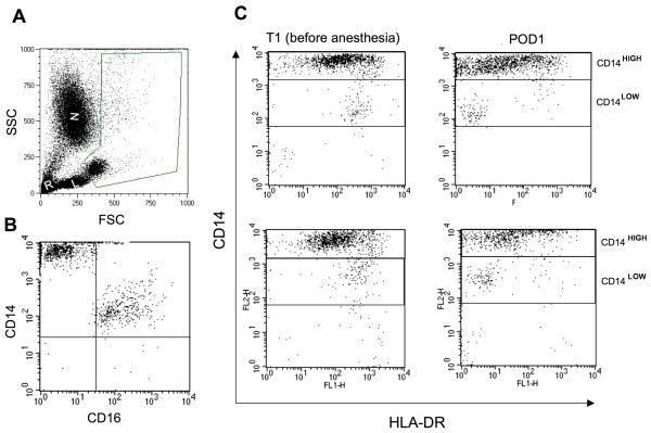
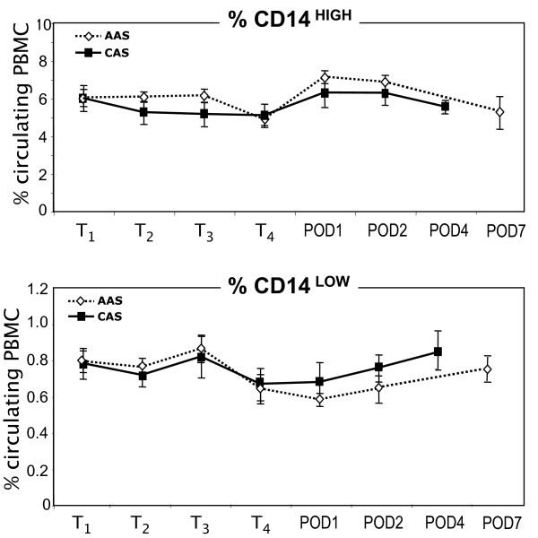
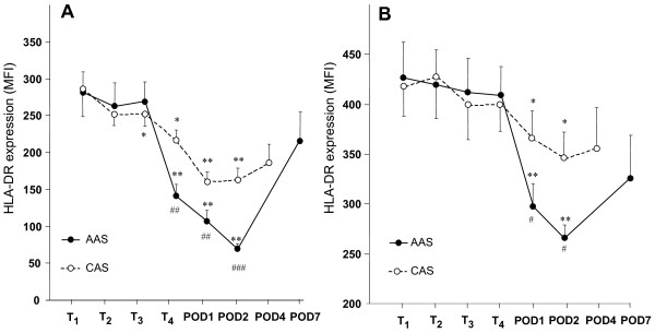
Methods: We studied this modification on CD14HIGH and CD14LOW monocytes of 20 SIRS patients undergoing abdominal aortic surgery (AAS), 20 patients undergoing carotid artery surgery (CAS), and 9 healthy controls, and we investigated mediators and intracellular molecules that may be involved in this process.

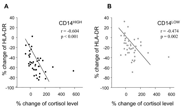
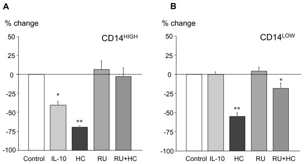
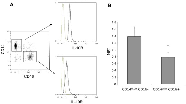
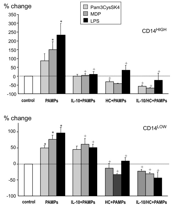
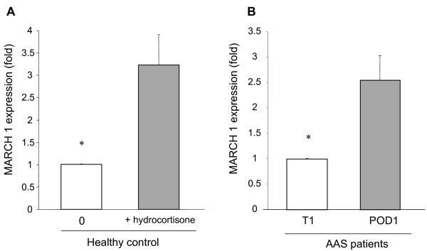
Results: HLA-DR on CD14HIGH monocytes started to decrease during surgery, after blood reperfusion, and was further reduced post-surgery. In contrast, HLA-DR expression on CD14LOW cells only decreased after surgery, and to a lesser extent than on CD14HIGH monocytes. Negative correlations were found between the reduction of HLA-DR expression and the change in cortisol levels for both subpopulations, whereas a negative correlation between interleukin-10 (IL-10) levels and HLA-DR modulation was only observed for CD14HIGH cells. In accordance with these ex vivo results, HLA-DR on CD14HIGH and CD14LOW monocytes of healthy donors was reduced following incubation with hydrocortisone, whereas IL-10 only acted on CD14HIGH subpopulation. Furthermore, flow cytometry revealed that the expression of IL-10 receptor was higher on CD14HIGH versus CD14LOW monocytes. In addition, hydrocortisone, and to a lesser extent IL-10, reversed the up-regulation of HLA-DR induced by bacterial products. Finally, membrane-associated RING-CH-1 protein (MARCH1) mRNA, a negative regulator of MHC class II, was up-regulated in monocytes of AAS patients on Day 1 post-surgery, and in those of healthy subjects exposed to hydrocortisone.

Conclusions: This study reveals that HLA-DR expression is modulated differently on CD14HIGH (classical) versus CD14LOW (inflammatory) monocytes after systemic inflammation.


Figures

Figure 1
Figure 1
Representative flow cytometry analysis of HLA-DR expression on CD14HIGH and CD14LOW monocyte subsets. Representative flow cytometry analysis of HLA-DR expression on CD14HIGH and CD14LOW monocyte subsets during and after surgery. (A) Flow cytometric analysis was performed on whole blood samples after elimination of neutrophils (N), lymphocytes (L) and red cells (R) using the forward scatter (FSC) and side scatter (SSC) characteristics. (B) A representative dot-plot showing that CD14HIGH monocytes were CD16- and CD14LOW monocytes were CD16+ in a patient before surgery. (C) Monocytes were analyzed using an anti-CD14 antibody coupled with phycoerythrin (PE) and an anti-HLA-DR antibody coupled with fluorescein isothiocyanate (FITC). Representative flow cytometric analysis for HLA-DR expression on CD14HIGH and CD14LOW monocytes performed before anesthesia (T1) and on postoperative Day 1 (POD1) are shown for two abdominal aortic surgery (AAS) patients.
Figure 2
Figure 2
Survey of CD14-positive subsets during and after surgery. Percent of CD14HIGH and CD14LOW subsets was followed in abdominal aortic surgery (AAS, close symbol) and carotid surgery (CAS, open symbol) patients, at T1 (before anesthesia), T2 (before incision), T3 (before clamping), T4 (after reperfusion), and on post-operative day 1, 2, 4 and 7 (POD1, 2, 4, 7). The results are expressed as percent of CD14-positive cells among leukocytes.
Figure 3
Figure 3
Monitoring of HLA-DR expression on CD14HIGH and CD14LOW monocyte subsets during and after surgery. Mean ± SEM corresponding to the mean fluorescence intensity (MFI) for HLA-DR expression on CD14HIGH (A) and CD14LOW (B) monocytes from patients undergoing abdominal aortic surgery (AAS, close symbol) or carotid surgery (CAS, open symbol) at T1 (before anesthesia), T2 (before incision), T3 (before clamping), T4 (after reperfusion), and on post-operative Days 1, 2, 4 and 7 (POD1, 2, 4, 7). The two curves in both panel A and B are significantly different (P = 0.001). * P < 0.05, ** P < 0.001 compared with the initial value (T1) for each monocyte subset. # P < 0.05, ## P < 0.01 and ### P < 0.001 when comparing the two groups of patients at one given time point.
Figure 4
Figure 4
Correlation between percent change in plasma cortisol levels and in HLA-DR expression on CD14HIGH and CD14LOW monocytes. Percent change was calculated between the levels observed before anesthesia (T1) and on postoperative Day 1 (POD1). Analysis with Spearman's rho correlation coefficient was performed for all 40 patients (AAS and CAS). (A) CD14HIGH monocytes; (B) CD14LOW monocytes.
Figure 5
Figure 5
Modulation of HLA-DR expression on CD14HIGH and CD14LOW monocytes by IL-10 or hydrocortisone. Blood samples from healthy donors were incubated for 24 hours without or with IL-10 (10 ng/ml), hydrocortisone (HC, 100 μM), and/or RU486 (20 μM), an antagonist of the glucocorticoid receptor. For both subpopulations, the results are expressed as percent change of mean fluorescence intensity for HLA-DR measured in untreated cells (control). The results are the mean ± SEM of five independent experiments with different donors. * P < 0.05, ** P < 0.01 compared with control by Mann-Whitney U-test.
Figure 6
Figure 6
Expression of the receptor for IL-10 (IL-10R) on CD14HIGH and CD14LOW monocytes. Flow cytometric analysis was performed on whole blood from healthy volunteers after elimination of polymorphonuclear cells, red cells and debris, using the forward scatter (FSC) and side scatter (SSC) characteristics. (A) CD14HIGHCD16- and CD14LOWCD16+ monocytes were analyzed for the expression of IL-10R. (B) The mean fluorescence intensity (MFI) for each subset is shown. The results are the mean ± SEM of seven independent experiments with different donors (* P < 0.05 using the Wilcoxon signed-rank test).
Figure 7
Figure 7
Modulation of HLA-DR expression on CD14HIGH and CD14LOW monocytes by different neuromediators, cytokines, PGE2 and ACTH. Blood samples from healthy donors were incubated for 24 hours without or with norepinephrine (NE, 10 nM), acetylcholine (AC, 10 μM), vasoactive intestinal peptide (VIP, 10 nM), pituitary adenylate cyclase-activating polypeptide (PACAP, 10 nM), substance P (SP, 1 μM), enkephalin (Enk, 1 μM), a mixture of all neuromediators, prostaglandin E2 (PGE2, 1 μM), adrenocorticotropin hormone (ACTH, 10 nM), transforming growth factor-β (TGF-β, 10 ng/ml), or tumor necrosis factor-α (TNF-α, 10 ng/ml). For both subpopulations, the results are expressed as the percent change of mean fluorescence intensity compared to HLA-DR measured in control cells. The results are the mean ± SEM of five independent experiments with different donors (* P < 0.05).
Figure 8
Figure 8
Modulation of HLA-DR expression on CD14HIGH and CD14LOW monocytes. Modulation of HLA-DR expression on CD14HIGH and CD14LOW monocytes by different PAMPs in the absence or presence of IL-10 and/or hydrocortisone. Blood samples from healthy donors were incubated for 24 hours without or with Pam3CysSK4 (100 ng/ml), muramyl dipeptide (MDP, 100 nM) or Escherichia coli LPS (100 ng/ml) in the absence or presence of IL-10 (10 ng/ml) and/or hydrocortisone (HC, 100 μM). For both subpopulations, the results are expressed as the percent change of mean fluorescence intensity compared to HLA-DR measured in untreated cells (control). The results are the mean ± SEM of four independent experiments with different donors. *P < 0.05, comparison between control and PAMPs --induced modulation. +P < 0.05, comparison between PAMPs alone and PAMPs + IL-10 and/or HC.
Figure 9
Figure 9
Increased expression of MARCH1 in monocytes after treatment. Increased expression of MARCH1 in monocytes after treatment with hydrocortisone in vitro or after abdominal aortic surgery. (A) MARCH1 mRNA expression in monocytes from healthy donors after a 24-hour incubation at 37°C in whole blood in the absence or presence of hydrocortisone (100 μM). MARCH1 expression was analyzed by qPCR and normalized as compared to GAPDH. The results are expressed as fold increase compared to untreated whole blood, and represent the mean ± SEM of six different donors. (B) MARCH1 mRNA expression in monocytes from AAS patients before anesthesia (T1) and one day post surgery (POD1). MARCH1 expression was analyzed by qPCR and normalized against that of GAPDH. The results are expressed as fold increase compared to the expression observed before surgery on T1, and represent the mean ± SEM of seven different patients. * P < 0.05 using the Wilcoxon signed-rank test.

References

    1. Bone RC, Grodzin CJ, Balk RA. Sepsis: A new hypothesis for pathogenesis of the disease process. Chest. 1997;112:235–243. doi: 10.1378/chest.112.1.235. - DOI - PubMed
    1. Adib-Conquy M, Cavaillon JM. Compensatory anti-inflammatory response syndrome. Thromb Haemost. 2009;101:36–47. - PubMed
    1. Venet F, Chung CS, Kherouf H, Geeraert A, Malcus C, Poitevin F, Bohe J, Lepape A, Ayala A, Monneret G. Increased circulating regulatory T cells (CD4(+)CD25 (+)CD127 (-)) contribute to lymphocyte anergy in septic shock patients. Intensive Care Med. 2009;35:678–686. doi: 10.1007/s00134-008-1337-8. - DOI - PMC - PubMed
    1. Muñoz C, Carlet J, Fitting C, Misset B, Bleriot JP, Cavaillon JM. Dysregulation of in vitro cytokine production by monocytes during sepsis. J Clin Invest. 1991;88:1747–1754. doi: 10.1172/JCI115493. - DOI - PMC - PubMed
    1. McCall CE, Grosso-Wilmoth LM, LaRue K, Guzman RN, Cousart SL. Tolerance to endotoxin-induced expression of the interleukin-1β gene in blood neutrophils of humans with the sepsis syndrome. J Clin Invest. 1993;91:853–861. doi: 10.1172/JCI116306. - DOI - PMC - PubMed

Publication types

MeSH terms

Substances