. 2010 Apr 9;38(1):41-53.
doi: 10.1016/j.molcel.2010.01.042.

Highly compacted chromatin formed in vitro reflects the dynamics of transcription activation in vivo

Affiliations

Highly compacted chromatin formed in vitro reflects the dynamics of transcription activation in vivo

Guohong Li et al. Mol Cell. .

Abstract

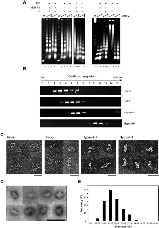
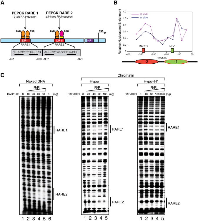
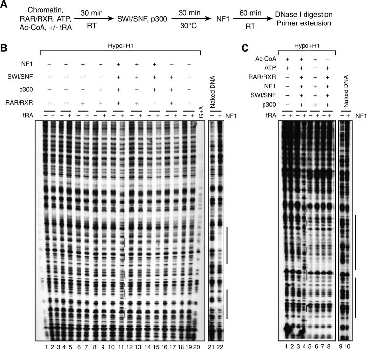
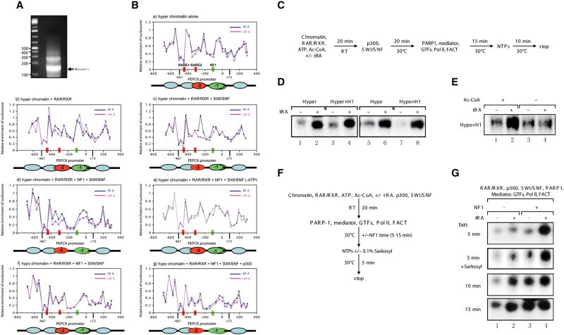
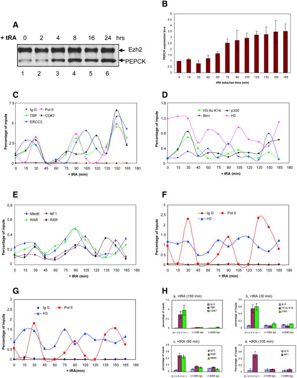
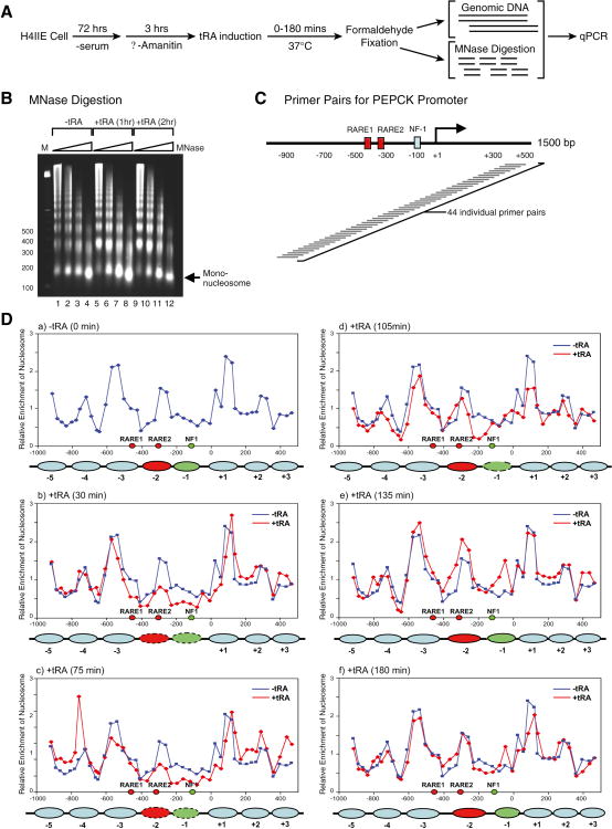
High-order chromatin was reconstituted in vitro. This species reflects the criteria associated with transcriptional regulation in vivo. Histone H1 was determinant to formation of condensed structures, with deacetylated histones giving rise to highly compacted chromatin that approximated 30 nm fibers as evidenced by electron microscopy. Using the PEPCK promoter, we validated the integrity of these templates that were refractory to transcription by attaining transcription through the progressive action of the pertinent factors. The retinoic acid receptor binds to highly compacted chromatin, but the NF1 transcription factor binds only after histone acetylation by p300 and SWI/SNF-mediated nucleosome mobilization, reflecting the in vivo case. Mapping studies revealed the same pattern of nucleosomal repositioning on the PEPCK promoter in vitro and in vivo, correlating with NF1 binding and transcription. The reconstitution of such highly compacted "30 nm" chromatin that mimics in vivo characteristics should advance studies of its conversion to a transcriptionally active form.


Figures

Figure 1
Figure 1
The reconstitution of 30-nm chromatin fiber in vitro: requirements for H1 incorporation and deacetylation of core histones. (A) Microccocal nuclease digestion analysis of chromatin assembled with supercoiled 5 kb template (pG5MLP 5S) using RSF and/or NAP-1 as a function of the presence of histone H1 either during (lane1-12) or after (lane 13-20) the chromatin assembly reaction. (B) Purification of reconstituted chromatin by sucrose gradient sedimentation. The fractions were analyzed by agarose gel electrophoresis and visualized using ethidium bromide staining. (C) Sucrose gradient peak fractions were subjected to analysis by electron microscopy (EM) using tungsten shadowing. Scale bar: 100 nm. (D) Gallery of side views of negatively stained compacted chromatin fibers in the presence of 0.5 mM MgCl2. Scale bar: 100 nm. Negative stain images are more appropriate for establishing the diameter of filaments as metal buildup around the particles in rotary shadowed images significantly increases their apparent diameter. (E) Bar graph representation of the diameters of chromatin fibers formed with hypoacetylated histones and histone H1 in the presence of 0.5 mM MgCl2 shown in Figure 1D. See also Figure S1.
Figure 2
Figure 2
The effects of chromatin structure on RAR/RXR binding to chromatin templates in vitro. (A) Diagram of the p(PEPCK-500)G template containing the PEPCK promoter used for in vitro DNase I footprinting and transcription assays. (B) Line graph of nucleosomal positioning at the PEPCK promoter in vivo (red) and in vitro (blue). The nucleosomes covering RARE2 and the NF1 binding site are represented as ovals in red and green, respectively. The mapping of nucleosomal positioning at the PEPCK promoter in vitro and in vivo is described in Experimental Procedures. Nucleosomal positioning in vivo shown here represents the nucleosome structure after tRA treatment (165 min). (C) DNase I footprinting analysis of RAR/RXR bound to the PEPCK promoter on naked DNA (left panel), or chromatin fiber assembled with either hyperacetylated histones (middle panel), or hypoacetylated histones and histone H1 (right panel). The binding sites for RAR/RXR (RARE1 and -2) are indicated by bond bars. See also Figure S2.
Figure 3
Figure 3
Remodeling/opening of 30-nm chromatin fiber in vitro: requirements for SWI/SNF and p300. (A) Reaction scheme for analyzing NF1 binding to compacted chromatin template p(PEPCK-500)G. (B, C) DNase I footprinting analysis for NF1 binding using chromatinized template assembled with hypoacetylated histones and histone H1, as a function of the presence of the factors indicated at the top and also shown in the reaction scheme in panel (A). (B) The binding site of NF1 is indicated by a bond bar, and hypersensitive sites are indicated by red stars. Naked DNA is shown as control. (C) ATP and Ac-CoA are required for the remodeling/opening of compacted chromatin mediated by SWI/SNF and p300 to facilitate NF1 binding. See also Figure S3.
Figure 4
Figure 4
Retinoid acid receptor (RAR/RXR)-induced chromatin transitions allow NF1 to facilitate ligand-dependent transcription at the PEPCK promoter in vitro. (A) Mononucleosomes derived from microccocal nuclease digestion of chromatinized template p(PEPCK-500)G assembled with hyperacetylated histone octamers using the RSF/NAP-1 system in vitro. (B) Nucleosomal mapping of the PEPCK promoter region within chromatinized p(PEPCK-500)G template assembled with hyperacetylated histone octamers (a-e) or hypoacetylated histone octamers (f, g) as a function of the presence of the factors indicated at the top of each panel. (C) The reaction scheme of the reconstituted transcription assays for (D) and (E). (D)In vitro reconstituted ligand-dependent transcription assays using the different chromatin templates indicated. (E) Optimal transcription from 30-nm chromatin fiber by RNAPII requires acetylation of core histones. (F) The reaction scheme of the reconstituted transcription assay for (G). (G) NF1 stimulates ligand-dependent transcription by RNAPII from compacted 30-nm chromatin fiber in the absence and presence of 0.1% Sarkosyl.
Figure 5
Figure 5
In vivo ChIP on the endogenous PEPCK promoter: cyclical and ordered recruitment of factors. (A) Western blot analysis of PEPCK expression upon tRA induction. (B) RT-qPCR analysis of PEPCK mRNA levels upon tRA induction. Values are the mean ± SD (standard deviation) of three independent experiments. (C, D, E)In vivo kinetic ChIP analyses of RNAPII and general transcriptional factors (C), coactivators and modified histones (D), and transcriptional activators and Mediator (E) at the PEPCK promoter. The amount of immunoprecipitated PEPCK promoter was quantified by q-PCR and normalized to the input DNA. (F-G)In vivo ChIP analyses of RNAPII and histone H3 at the PEPCK coding regions: +1000 nt (F) and +2000 nt (G) from the transcription start site. (H) Enrichment of general transcriptional factors (a), coactivators and modified histones (b), transcriptional activators and Mediator (c-d) at the PEPCK promoter, but not at the coding regions. For simplicity, a representative time point is shown in each case, but similar results were obtained at all other 15 min increments of time. Percent of input are the mean ± SD (standard deviation) of three independent experiments.
Figure 6
Figure 6
Dynamic nucleosome repositioning at the PEPCK promoter during transcription activation in vivo. (A) Protocol used for nucleosomal mapping in vivo. (B) Microccocal nuclease digestion of chromatin isolated from H4IIE cells as a function of treatment with tRA. (C) A schematic of the tiling primers covering the PEPCK promoter used for identifying in vivo nucleosomal positions are shown. (D) The dynamic change in nucleosomal positions at the PEPCK promoter in the absence (blue graph) and in the presence (red graph) of tRA induction (30-180 min). The filled ovals represent the nucleosomes covering RARE2 (red) and NF1 (green) that are repositioned upon tRA exposure.
Figure 7
Figure 7
Model for the dynamic folding and opening of 30 nm chromatin fiber for RNAPII transcription. See text for details.

References

    1. Aoyagi S, Archer TK. Dynamics of coactivator recruitment and chromatin modifications during nuclear receptor mediated transcription. Mol Cell Endocrinol. 2008;280:1–5. - PMC - PubMed
    1. Archer TK, Lefebvre P, Wolford RG, Hager GL. Transcription factor loading on the MMTV promoter: a bimodal mechanism for promoter activation. Science. 1992;255:1573–1576. - PubMed
    1. Belikov S, Astrand C, Holmqvist PH, Wrange O. Chromatin-mediated restriction of nuclear factor 1/CTF binding in a repressed and hormone-activated promoter in vivo. Mol Cell Biol. 2004;24:3036–3047. - PMC - PubMed
    1. Belikov S, Gelius B, Almouzni G, Wrange O. Hormone activation induces nucleosome positioning in vivo. EMBO J. 2000;19:1023–1033. - PMC - PubMed
    1. Bruno M, Flaus A, Stockdale C, Rencurel C, Ferreira H, Owen-Hughes T. Histone H2A/H2B dimer exchange by ATP-dependent chromatin remodeling activities. Mol Cell. 2003;12:1599–1606. - PMC - PubMed

Publication types

MeSH terms