Comparative Study
. 2010 Aug 1;80(3):325-34.
doi: 10.1016/j.bcp.2010.04.007. Epub 2010 Apr 10.

Transcriptional up-regulation of SOD1 by CEBPD: a potential target for cisplatin resistant human urothelial carcinoma cells

Affiliations
Comparative Study

Transcriptional up-regulation of SOD1 by CEBPD: a potential target for cisplatin resistant human urothelial carcinoma cells

Tzyh-Chyuan Hour et al. Biochem Pharmacol. .

Abstract

Bladder cancer is the fourth most common type of cancer in men (ninth in women) in the United States. Cisplatin is an effective agent against the most common subtype, urothelial carcinoma. However, the development of chemotherapy resistance is a severe clinical problem for the successful treatment of this and other cancers. A better understanding of the cellular and molecular events in response to cisplatin treatment and the development of resistance are critical to improve the therapeutic options for patients. Here, we report that expression of the CCAAT/enhancer binding protein delta (CEBPD, C/EBPdelta, NF-IL6beta) is induced by cisplatin in the human bladder urothelial carcinoma NTUB1 cell line and is specifically elevated in a cisplatin resistant subline. Expression of CEBPD reduced cisplatin-induced reactive oxygen species (ROS) and apoptosis in NTUB1 cells by inducing the expression of Cu/Zn-superoxide dismutase (SOD1) via direct promoter transactivation. Several reports have implicated CEBPD as a tumor suppressor gene. This study reveals a novel role for CEBPD in conferring drug resistance, suggesting that it can also be pro-oncogenic. Furthermore, our data suggest that SOD inhibitors, which are already used as anti-angiogenic agents, may be suitable for combinatorial chemotherapy to prevent or treat cisplatin resistance in bladder and possibly other cancers.


Conflict of interest statement

Disclosure of potential conflicts of interest

No potential conflicts of interest were disclosed.

Figures
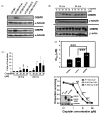
Fig 1
Fig 1
CEBPD protein is expressed specifically in a cisplatin resistant subline and over-expressing CEBPD enhances cisplatin resistance. (A) Western blot analysis of CEBPD and CEBPB protein expression in NTUB1 and drug resistant sublines (NTUB1/P(14), NTUB1/G(1.5), NTUB1/As(1.5) and NTUB1/T(0.017)). Expression of α-tubulin was used as a loading control. (B) NTUB1 cells were treated with 0, 10, 20, 30 or 40 μM of cisplatin and the proteins were harvested at 12 or 24 h as indicated. Western blot analysis was performed with antibodies for CEBPD, α-tubulin, CEBPB and β-actin. (C) Q-RT-PCR analysis of CEBPD mRNA expression from the same treatment as (B). CEBPD mRNA expression normalized to cyclophilin A expression is shown as fold over untreated samples at 12 h. Data represent means ± standard deviation (SD) of five independent samples, each analyzed in duplicate (***P < 0.005 and *P < 0.05). (D) IC50 values of transient transfected cells were performed by MTT assay as described in Section 2 (***P < 0.005). Data represent means ± standard deviation (SD) of three independent experiments. (E) A representative MTT assay of established CEBPD expressing clones and the IC50 values of each group were calculated by the median-effect analysis and presented as means ± SD with ***P < 0.005 compared to N-V and N-ΔDBD.
Fig 2
Fig 2
The DNA-binding activity of CEBPD is essential for the inhibition of cisplatin-induced apoptosis. (A) Representative images of N-V, N-D#1 and N-ΔDBD clones, seeded onto glass coverslips and treated with 0, 10, or 20 μM of cisplatin for 24 h. The nuclei were stained with DAPI and photographed by microscopy. The arrows point to apoptotic cells. (B) The indicated cells were treated with 0, 10, or 20 μM of cisplatin for 24 h and the cell cycle profile of each sample was determined by flow cytometry. The means ± SD of 2–4 independent experiments is shown with *P < 0.05 comparing N-D#1 and N-V at 20 μM cisplatin. (C) A representative flow cytometry of ROS in untreated N-V, N-D#1 and N-ΔDBD cells. The M1 region is indicated. (D) The N-V, N-D#1 and N-R198A cells were treated with 0 or 20 μM of cisplatin for 24 h. The ROS levels were determined and transformed to M1 ratios as described in Section 2. The data represent the percentage of ROS production efficiency compared to the N-V untreated samples (means ± SD) of four independent experiments with ***P < 0.005.
Fig 3
Fig 3
CEBPD expression is positively correlated with SOD1 protein levels. (A) Western blot analysis of SOD1 and PRX protein expression in NTUB1 stable clones. α-tubulin was used as a control for protein loading. (B) N-V and N-D#1 cells were treated with 20 μM cisplatin for 24 h followed by Western analysis of CEBPD and SOD1 expression. β-Actin was used as a control for protein loading. (C) Cisplatin resistant subline NTUB1/P14 was transiently transfected with shRNA expression constructs directed against GFP (GFPsi) as negative control, CEBPD (CEBPDsi), or SOD1 (SODsi). CEBPD and SOD1 expression were assessed 24 h later compared to β-actin as control.
Fig 4
Fig 4
SOD1 is a target gene of CEBPD. (A) Scheme of the human SOD1 promoter from positions +74 to −1499 bp. The potential C/EBP binding sites were predicted by the program “TFSEARCH” [45]. Five predicted C/EBP binding sites (filled ovals) are shown at positions −1321 to −1311, −723 to −713, −707 to −697, −694 to 684 and −248 to −241 bp. The primers (arrows) and PCR product (bar) used for ChIP (panel D) are indicated. (B) Luciferase reporter activity from 0.1 μg of SOD1 luciferase reporter plasmid transiently co-transfected with 0.1, 0.2 or 0.3 μg of CEBPD expression plasmid in NTUB1 cells. The results were normalized by total protein concentration and are represented relative to vector control (means ± SD from three independent experiments performed in duplicate each) with ***P < 0.005. (C) Luciferase reporter activity from 0.1 μg of SOD1 luciferase reporter plasmid transiently co-transfected with 0.3 μg of vector (V), WT CEBPD (WT), or R198A mutant (R198A) CEBPD expression plasmids in NTUB1 cells. Shown are the means ± SD from at least three independent experiments performed in duplicate, relative to vector transfected cells with *P < 0.05 and **P < 0.01. (D) NTUB1 cells were treated with 0 or 20 μM cisplatin for 24 h. The sonicated chromatin was subjected to ChIP analysis by CEBPD antibody or control IgG. The amplified DNA products were resolved by agarose gel electrophoresis, stained with ethidium bromide and photographed. The SOD1 promoter-specific PCR product is indicated by the arrow. (E) SOD1 mRNA expression was examined by Q-RT-PCR in NTUB1 cells treated with 20 μM cisplatin for 24 h and NTUB1/P(14) cells compared to untreated NTUB1 (Results are shown as fold of change to NTUB1 cells in mean ± SD, from three independent experiments performed in duplicate each; ***P < 0.005).
Fig 5
Fig 5
SOD1 inhibition potentiates cisplatin cytotoxicity. (A) Representative flow cytometry of ROS levels in NTUB1 cells treated for 24 h with 20 μM cisplatin and/or 20 mM TETA. (B) NTUB1 and (C) NTUB1/P(14) cells were subjected to cytotoxicity analysis with combinations of cisplatin and TETA (0.5, 2 and 5 mM) by MTT assay as described in Section 2. The percentage of cell survival relative to untreated cells is presented. ***P < 0.005 and *P < 0.05 between cisplatin alone and combined samples, respectively.
Fig 6
Fig 6
Model for CEBPD-mediated cisplatin resistance. Cisplatin causes DNA damage as well as reactive oxygen species (ROS), which trigger cell cycle arrest or/and apoptosis. Cisplatin induces CEBPD by an as of yet unidentified mechanism which directly activates superoxide dismutase (SOD1) gene expression. Superoxide anion (O2•−) is dismutated by SOD1 and converted to H2O2 which can be further neutralized to water and oxygen by catalase. The reduced ROS levels in NTUB1 cells cause the cisplatin resistant phenotype. The inhibition of SOD1 by triethylenetetramine (TETA) potentiates the cisplatin cytotoxicity and abrogates cisplatin resistance.

References

    1. Bischoff CJ, Clark PE. Bladder cancer. Curr Opin Oncol. 2009;21:272–7. - PubMed
    1. Kaufman DS. Challenges in the treatment of bladder cancer. Ann Oncol. 2006;17(Suppl 5):v106–12. - PubMed
    1. Bellmunt J, Albiol S, Suarez C, Albanell J. Optimizing therapeutic strategies in advanced bladder cancer: update on chemotherapy and the role of targeted agents. Crit Rev Oncol Hematol. 2009;69:211–22. - PubMed
    1. Zamble DB, Lippard SJ. Cisplatin and DNA repair in cancer chemotherapy. Trends Biochem Sci. 1995;20:435–9. - PubMed
    1. Sedletska Y, Giraud-Panis MJ, Malinge JM. Cisplatin is a DNA-damaging anti-tumour compound triggering multifactorial biochemical responses in cancer cells: importance of apoptotic pathways. Curr Med Chem. 2005;5:251–65. - PubMed

Publication types

MeSH terms