

An in vitro assay system for studying synapse formation between nociceptive dorsal root ganglion and dorsal horn neurons
- PMID: 20385165
- PMCID: PMC2880384
- DOI: 10.1016/j.jneumeth.2010.04.002
An in vitro assay system for studying synapse formation between nociceptive dorsal root ganglion and dorsal horn neurons
Abstract
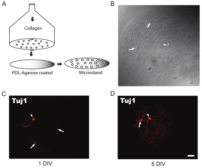
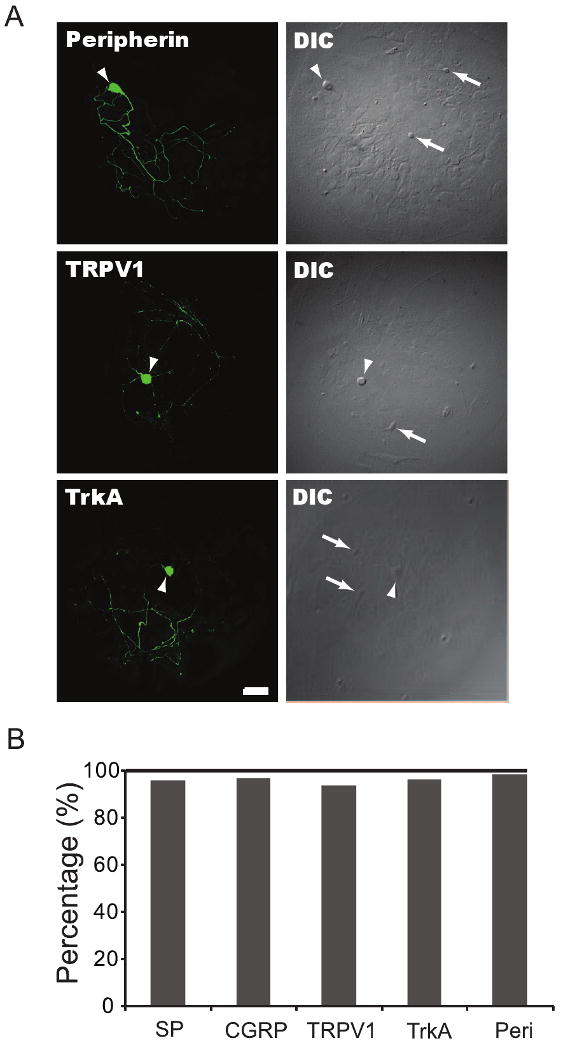
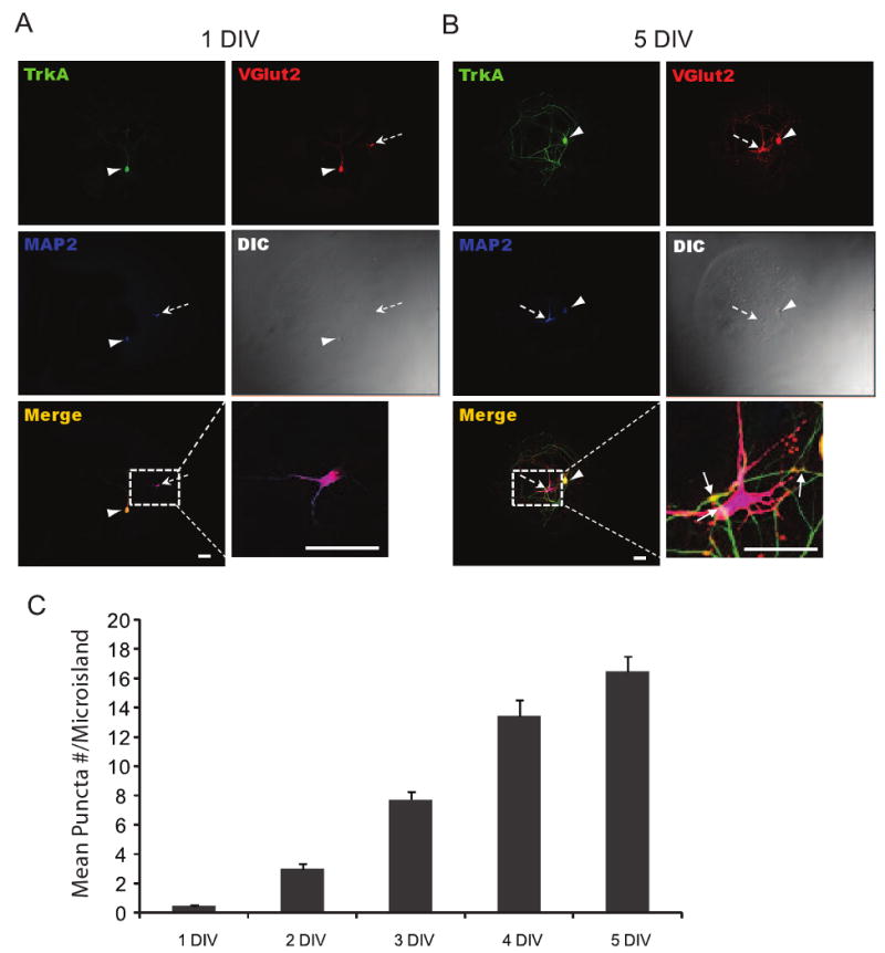
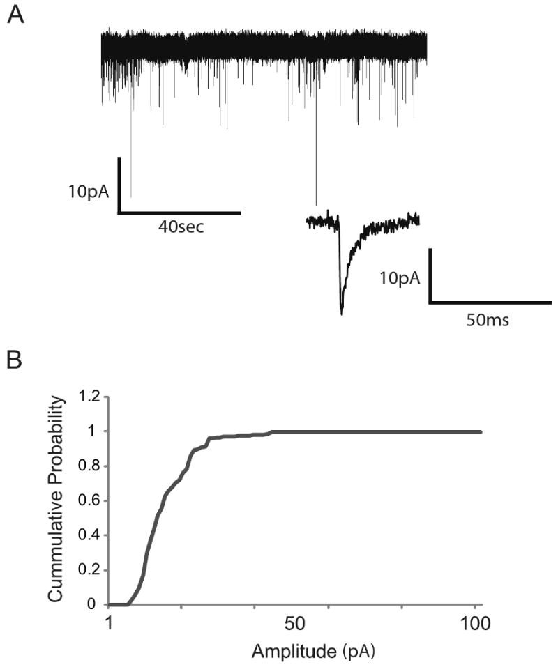
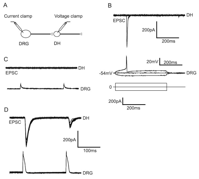
Synapses between nociceptive dorsal root ganglion (DRG) neurons and spinal cord dorsal horn neurons represent the first loci for transmission of painful stimuli. Our knowledge of the molecular organization and development of these synapses is sparse due, partly, to a lack of a reliable model system that reconstitutes synaptogenesis between these two neuronal populations. To address this issue, we have established an in vitro assay system consisting of separately purified DRG neurons and dorsal horn neurons on astrocyte microislands. Using immunocytochemistry, we have found that 97%, 93%, 98%, 96%, and 94% of DRG neurons on these microislands express markers often associated with nociceptive neurons including Substance P, TRPV1, calcitonin-gene related peptide (CGRP), TrKA, and peripherin, respectively. Triple labeling with these nociceptive-like markers, synaptic vesicle marker Vglut2 and using MAP2 as a dendritic marker revealed the presence of nociceptive-like markers at synaptic terminals. Using this immunocytochemical approach, we counted contact points as overlapping MAP2/Vglut2 puncta and showed that they increased with time in culture. Single and dual patch-clamp recordings showed that overlapping Vglut2/MAP2 puncta observed after a few days in culture are likely to be functional synapses between DRG and dorsal horn neurons in our in vitro assay system. Taken together, these data suggest our co-culture microisland model system consists of mostly nociceptive-like DRG neurons that express presynaptic markers and form functional synapses with their dorsal horn partners. Thus, this model system may have direct application for studies on factors regulating development of nociceptive DRG/dorsal horn synapses.
Copyright (c) 2010 Elsevier B.V. All rights reserved.
Figures
References
-
- Albuquerque C, Lee CJ, Jackson AC, MacDermott AB. Subpopulations of GABAergic and non-GABAergic rat dorsal horn neurons express Ca2+-permeable AMPA receptors. Eur J Neurosci. 1999;11:2758–2766. - PubMed
-
- Albuquerque C, Joseph DJ, Choudhury P, MacDermott AB. Preparation of coverslips for neuronal cultures. CSH Protoc. 2009 Aug 1;2009(8) pdb.prot5272. - PubMed
-
- Albuquerque C, Joseph DJ, Choudhury P, MacDermott AB. Dissection, plating, and maintenance of cortical astrocyte cultures. CSH Protoc. 2009 Aug 1;2009(8) pdb.prot5273. - PubMed
-
- Albuquerque C, Joseph DJ, Choudhury P, MacDermott AB. Dissection, plating, and maintenance of dorsal horn neuron cultures. CSH Protoc. 2009 Aug 1;2009(8) pdb.prot5274. - PubMed
-
- Albuquerque C, Joseph DJ, Choudhury P, MacDermott AB. Dissection, Plating and maintenance of dissociated dorsal root ganglion (DRG) neurons for mono-culture and for co-culture with dorsal horn neurons. CSH Protoc. 2009 Aug 1;2009(8) pdb.prot5275.
Publication types
MeSH terms
Substances
Grants and funding
LinkOut - more resources
Full Text Sources
Research Materials
