

Dysfunction of the magnocellular stream in Alzheimer's disease evaluated by pattern electroretinograms and visual evoked potentials
- PMID: 20385208
- PMCID: PMC3227554
- DOI: 10.1016/j.brainresbull.2010.04.001
Dysfunction of the magnocellular stream in Alzheimer's disease evaluated by pattern electroretinograms and visual evoked potentials
Abstract
Background: Visuo-spatial disturbances could represent a clinical feature of early stage Alzheimer's disease (AD). The magnocellular (M) pathway has anatomo-physiological characteristic which make it more suitable for detecting form, motion and depth compared with parvocellular one (P).
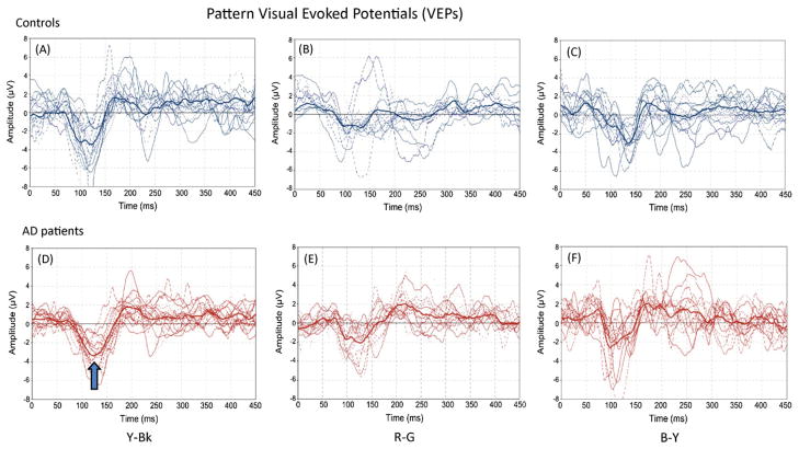
Objective: Aim of our study was to evaluate specific visual subsystem involvement in a group of AD patients, recording isoluminant chromatic and luminance pattern electroretinograms and pattern visual evoked potentials.
Material and methods: data were obtained from 15 AD patients (9 females and 6 males, mean age+/-1SD: 77.6+/-4.01 years) not yet undergoing any treatment, and from 10 age-matched healthy controls. Diagnosis of probable AD was clinically and neuroradiologically established. PERGs were recorded monocularly in response to equiluminant red-green (R-G), blue-yellow (B-Y) and luminance yellow-black (Y-Bk) horizontal square gratings of 0.3c/deg and 90% contrast, reversed at 1Hz. VEPs were recorded in response to full-field (14 deg) equiluminant chromatic R-G, B-Y and luminance Y-Bk sinusoidal gratings of 2c/deg, presented in onset (300ms)-offset (700ms) mode, at the contrast levels of 90%.
Results: All data were retrieved in terms of peak-amplitude and latency and assessed using the Student's t-test for paired data. Temporal differences of PERGs and VEPs, evoked by Y-Bk grating in AD patients compared with controls, suggest a specific impairment of the magnocellular stream.
Conclusions: Our study support the hypothesis that the impairment of the PERGs and VEPs arising from the magnocellular streams of visual processing may indicate a primary dysfunction of the M-pathways in AD.
Published by Elsevier Inc.
Conflict of interest statement
We disclose any actual or potential conflict of interest including any financial, personal or other relationships with other people or organizations within three years of beginning our work submitted that could inappropriately influence it.
Figures

References
-
- Albert MS, Duffy FH, McAnulty GB. Electrophysiologic comparisons between two groups of patients with Alzheimer’s disease. Arch Neurol. 1990;47:857–863. - PubMed
-
- Aoki C, Kabak S. Cholinergic terminals in the cat visual cortex: ultrastructural basis for interaction with glutamate-immunoreactive neurons and other cells. Vis Neurosci. 1992;8:177–191. - PubMed
-
- Bayer AU, Ferrari F, Erb C. High occurrence rate of glaucoma among patients with Alzheimer’s disease. Eur Neurol. 2002;47:165–168. - PubMed
-
- Beelke M, Sannita WG. Cholinergic function and dysfunction in the visual system. Methods Find Exp Clin Pharmacol. 2002;24(Suppl D):113–117. - PubMed
-
- Berisha F, Feke GT, Trempe CL, McMeel JW, Schepens CL. Retinal abnormalities in early Alzheimer’s disease. Invest Ophthalmol Vis Sci. 2007;48:2285–2289. - PubMed
Publication types
MeSH terms
Grants and funding
LinkOut - more resources
Full Text Sources
Other Literature Sources
Medical
