. 2010 Apr 13;17(4):319-32.
doi: 10.1016/j.ccr.2010.02.030.

ERbeta impedes prostate cancer EMT by destabilizing HIF-1alpha and inhibiting VEGF-mediated snail nuclear localization: implications for Gleason grading

Affiliations

ERbeta impedes prostate cancer EMT by destabilizing HIF-1alpha and inhibiting VEGF-mediated snail nuclear localization: implications for Gleason grading

Paul Mak et al. Cancer Cell. .

Erratum in

  • Cancer Cell. 2010 Jun 15;17(6):622

Abstract

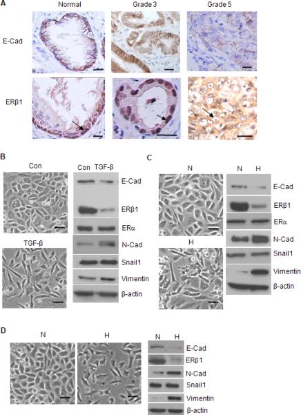
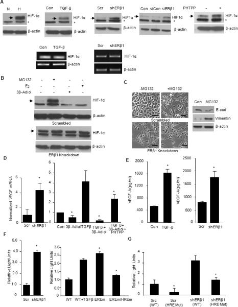
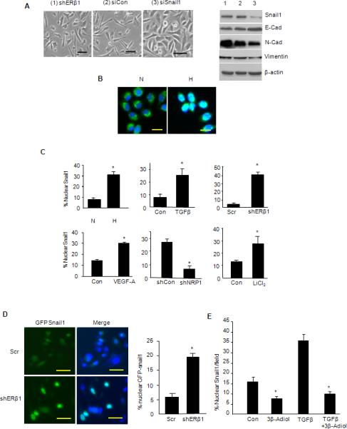
High Gleason grade prostate carcinomas are aggressive, poorly differentiated tumors that exhibit diminished estrogen receptor beta (ERbeta) expression. We report that a key function of ERbeta and its specific ligand 5alpha-androstane-3beta,17beta-diol (3beta-adiol) is to maintain an epithelial phenotype and repress mesenchymal characteristics in prostate carcinoma. Stimuli (TGF-beta and hypoxia) that induce an epithelial-mesenchymal transition (EMT) diminish ERbeta expression, and loss of ERbeta is sufficient to promote an EMT. The mechanism involves ERbeta-mediated destabilization of HIF-1alpha and transcriptional repression of VEGF-A. The VEGF-A receptor neuropilin-1 drives the EMT by promoting Snail1 nuclear localization. Importantly, this mechanism is manifested in high Gleason grade cancers, which exhibit significantly more HIF-1alpha and VEGF expression, and Snail1 nuclear localization compared to low Gleason grade cancers.


Figures

Figure 1
Figure 1. ERβ1 and EMT of PCa
(A) Specimens of normal glandular epithelium, Gleason grade 3 and 5 PCa were stained for E-cadherin and ERβ1 and photographed. ERβ1 is localized in the nuclei of basal cells in the normal prostate and nuclei of grade 3 tumor cells (arrow). In contrast, nuclear ERβ1 staining is absent in grade 5 PCa (arrow). The data are representative of 3 separate specimens for each classification. Scale bars = 20 μm. (B) PC3 cells were treated with PBS (con) or TGF-β for 3 days, photographed and extracts were analyzed for the expression of EMT markers and ERβ1 by immunoblotting. PC3 (C) or LNCaP (D) cells were maintained in either normoxia (N) or hypoxia (H) (0.5% O2) for 24 hrs, photographed and extracts from these cells were immunoblotted as described above. Scale bars = 50 μm. See also Figure S1.
Figure 2
Figure 2. ERβ1 Sustains An Epithelial Phenotype and Impedes EMT in PCa
(A) PC3 cells that express either an ERβ1 shRNA (shERβ1) or scrambled shRNA (shCon) or parental cells were photographed. (B) Extracts of these cells were immunoblotted for ERβ1 and EMT markers. (C) Extracts of PC3 cells that express either an ERα siRNA (siERα) or a scrambled siRNA (Scr) were immunoblotted for E-cadherin, N-cadherin, ERα and ERβ1. (D) The relative expression of E-cadherin and vimentin was assayed in PC3 cells that stably express either a control shRNA or ERβ1 shRNA by qPCR using PGK1 as an internal control. The data represent the average of 2 experiments. (E) PC3 cells expressing a scrambled shRNA (Scr) or an ERβ1 shRNA (shERβ1) were transfected with an E-cadherin promoter reporter construct (Src+ and shERβ1+) or pGL2 Basic vector as a control (Scr and shERβ1) and assayed for luciferase activity. The data represent the mean of Firefly luciferase activity normalized to Renilla from 3 separate experiments (±SEM) with P-value (*) <0.05. (F) PC3 cells that express either a control shRNA (shCon) or ERβ1 shRNA (shERβ1) were assessed for their ability to either migrate or invade. The data represent the mean of 3 separate experiments (±SEM) with P-value (*) <0.05. (G) PC3 cells transfected with either control siRNA (siCon) or ERβ1 SMARTpool siRNA (siERβ1) and the parental cells were examined for morphology and EMT marker expression after 3 days. Scale bars = 50 μm.
Figure 3
Figure 3. The ERβ1 Ligand 3β-Adiol Sustains An Epithelial Phenotype in PCa
PC3 cells were incubated with either DMSO (Con), the ERβ1 antagonist PHTPP (A) or 3β-Adiol (B) for 3 days, and morphology and EMT marker expression were examined. PC3 cells were treated with TGF-β in the absence or presence of estradiol-17β (E2) (10 nM) or 3β-Adiol (1 μM) and examined for morphology (C) and expression of ERβ1 and EMT markers (D: left panel). Cells treated in the absence or presence of TGF-β were also examined for ERβ1 transcripts by RT-PCR (D: right panel). ERβ1 knockdown cells (shERβ1) untreated or treated with either 3β-Adiol (1 μM) or E2 (10 nM) were examined for morphology and expression of EMT markers (E). Scale bars = 50 μm.
Figure 4
Figure 4. ERβ1 Destabilizes HIF-1α Protein and Represses HIF-1-mediated transcription of VEGF-A
(A) PC3 cells maintained in either normoxia (N) or hypoxia (H) for 24 hrs, treated with PBS (Con) or TGF-β, transfected with control or ERβ1 shRNA or siRNA, or treated with PHTPP were analyzed for the expression of HIF-1α by immunoblotting. * denotes a non-specific band. HIF-1α mRNA was detected by RT-PCR in TGF-β–stimulated cells and shRNA transfected cells. (B) PC3 cells (scrambled control cells) or ERβ1 knockdown cells were treated in the absence or presence of MG132 (1 μM), 3β-Adiol (1 μM) or E2 (10 nM) for 6 hours and immunoblotted for HIF-1α. (C) PC3 cells (scrambled control cells) or ERβ1 knockdown cells were treated in the absence or presence of MG132 (1 μM) for 6 hours and photographed. Scale bars = 50 μm. Extracts of the control cells treated with MG132 were immunoblotted for E-cadherin, vimentin and β-actin. (D) PC3 cells expressing a scrambled shRNA (Scr) or an ERβ1 shRNA (shERβ1) were analyzed for VEGF-A mRNA expression by qPCR (left graph). PC3 cells were treated with PBS (Con) or TGF-β in the absence or presence of 3β-Adiol (1 μM) or 3β-Adiol (1 μM) plus PHTPP (5 μM). After 3 days, cells were analyzed for VEGF-A mRNA expression by qPCR (right graph). (E) VEGF-A secretion in culture medium from PC3 cells treated with PBS (Con) or TGF-β or transfected with control or ERβ1 shRNAs was quantified by ELISA. (F) Scrambled control cells (Scr) or ERβ1 knockdown cells (shERβ1) were transfected with a VEGF promoter reporter construct and luciferase activity normalized to Renilla was measured (left graph). PC3 cells were transfected with a wild type VEGF promoter reporter construct in the absence (Wt) or presence of TGF-β (WT+TGFβ). Concurrently, cells were transfected with the reporter construct containing either a mutated ERE (EREm) or both a mutated ERE and HRE (EREm/HREm) and normalized luciferase activity was measured (right graph). (G) Scrambled control cells (Scr) or ERβ1 knockdown cells (shERβ) were transfected either with a wild-type HRE reporter construct: Scr (Wt) or shERβ (Wt) or with a mutated version of the HRE reporter construct: Scr (mut) or shERβ (mut) under hypoxic conditions for 16-18 hours and normalized luciferase activity was measured. All data are the mean of 3 separate experiments with SEM and P value (*) < 0.05 indicated.
Figure 5
Figure 5. VEGF-A Promotes EMT and the VEGF Receptor NRP1 is Necessary for EMT and Regulates GSK-3β Activity
(A) PC3 cells were grown in RPMI medium in the absence or presence of recombinant VEGF165 (50 ng/ml) for 24 hours. Cells were photographed and extracts were immunoblotted to assess expression of EMT markers. (B) Photomicrographs of PC3 cells that express either an empty vector (shCon), GFP shRNA (shGFP) or 2 different NRP1 shRNAs (shNRP1A and shNRP1B) were treated with or without TGF-β for 3 days. Extracts from these cells were immunoblotted for NRP1, as well as EMT markers. (C) Extracts from PC3 cells stimulated with either TGF-β, normoxia (N) or hypoxia (H), or ERβ1 knockdown cells (shERβ1) were immunoblotted with Abs specific for pAkt (Ser473), pGSK-3β (Ser9), Akt, GSK-3β and β-actin. (D) Extracts of LNCaP cells maintained in either normoxia (N) or hypoxia (H) for 24 hrs were immunoblotted with the same Abs. (E) PC3 cells were treated in the absence or presence of 3β-Adiol or PHTPP and subsequently analyzed for phospho-GSK3β, total GSK3β and Snail1 expression. Scale bars = 50 μm.
Figure 6
Figure 6. ERβ1 and EMT Regulate Snail1 Nuclear Localization
(A) PC3 cells that express an ERβ1 shRNA were transfected with either a control siRNA (siCon) or Snail1 siRNA (siSnail1), and analyzed for morphology and expression of EMT markers. (B) Snail1 was visualized by immunofluorescence microscopy in PC3 cells maintained in either normoxia (N) or hypoxia (H) for 48 hrs. The photomicrographs shown represent the merged images obtained from Snail1 staining (green; FITC) and nuclear staining (blue; DAPI). Note that in normoxia, Snail1 staining is predominantly cytoplasmic and excluded from nuclei. In hypoxia, however, Snail1 localization in nuclei is evidenced by ‘whitish-blue’ staining. (C) The percentage of nuclei that had Snail1 staining was quantified in PC3 cells maintained in normoxia (N) and hypoxia (H), and in cells stimulated with TGF-β or VEGF-A, as well as in PC3 cells in which ERβ1 or NRP1 expression was depleted by shRNA. Snail1 nuclear localization was also quantified in PC3 cells treated with LiCl2, a GSK-3β inhibitor. The data represent the mean of 3 separate experiments with SEM and P value (*) <0.05 indicated. (D) PC3 cells that express either a scrambled shRNA (Scr) or ERβ1 shRNA (shERβ1) were transfected with a GFP-Snail1 construct. GFP and DAP1 were visualized and the images merged. Note the nuclear localization of GFP-Snail1 as evidenced by the whitish blue staining that is associated with loss of ERβ1 expression. The bar graph represents the quantification of nuclear GFP-Snail1 from 3 independent experiments (±SEM) and P value (*) <0.05 indicated. (E) PC3 cells were treated with TGB-β in the absence or presence of 3β-Adiol (1 μM) and nuclear Snail1 was quantified. The data represent the mean of 3 separate experiments with SEM and P-value (*) <0.05 indicated.
Figure 7
Figure 7. HIF-1α/VEGF/Snail1 Pathway is Manifested in High Gleason Grade PCa
Thirty specimens of human PCa including 20 Gleason grade 3 tumors and 10 Gleason grade 5 tumors were immunostained for ERβ1 (A), HIF-1α (B), VEGF-A (C) and Snail1 (E). Semi-quantitative analysis of IHC staining was performed for all samples that assessed both the percentage of cells stained and the intensity of the staining, and this analysis is reported as the Quotient (Q) of these two parameters (±SD). The significance of the difference in Q between Gleason grade 3 and 5 as determined by Students t test is shown for each bar graph. Photomicrographs representative of the mean Q for each IHC staining are shown. (D) Microdissected samples from grade 3 and grade 5 PCa were analyzed for the expression of VEGF-A mRNA by qPCR and the data represent the average of 7 separate specimens for each grade. Red scale bars = 25 μm; black scale bars = 50 μm.
Figure 8
Figure 8. Proposed model for how ERβ1 sustains an epithelial phenotype and represses a mesenchymal phenotype
The interaction of ERβ1 with its ligand 3β-Adiol represses an EMT by destabilizing HIF-1α and inhibiting VEGF-A transcription. Stimuli that induce an EMT diminish ERβ1 expression resulting in increased VEGF-A expression and the consequent activation of a VEGF-A/NRP1 signaling pathway that inhibits GSK-3β and promotes Snail1 nuclear localization.

Comment in

References

    1. Acevedo VD, Gangula RD, Freeman KW, Li R, Zhang Y, Wang F, Ayala GE, Peterson LE, Ittmann M, Spencer DM. Inducible FGFR-1 activation leads to irreversible prostate adenocarcinoma and an epithelial-to-mesenchymal transition. Cancer Cell. 2007;12:559–571. - PubMed
    1. Andriole GL, Crawford ED, Grubb RL, 3rd, Buys SS, Chia D, Church TR, Fouad MN, Gelmann EP, Kvale PA, Reding DJ, et al. Mortality results from a randomized prostate-cancer screening trial. N Engl J Med. 2009;360:1310–1319. - PMC - PubMed
    1. Bachelder RE, Crago A, Chung J, Wendt MA, Shaw LM, Robinson G, Mercurio AM. Vascular endothelial growth factor is an autocrine survival factor for neuropilin-expressing breast carcinoma cells. Cancer Res. 2001;61:5736–5740. - PubMed
    1. Bachelder RE, Yoon SO, Franci C, de Herreros AG, Mercurio AM. Glycogen synthase kinase-3 is an endogenous inhibitor of Snail transcription: implications for the epithelial-mesenchymal transition. J Cell Biol. 2005;168:29–33. - PMC - PubMed
    1. Bae D, Lu S, Taglienti CA, Mercurio AM. Metabolic stress induces the lysosomal degradation of neuropilin-1 but not neuropilin-2. J Biol Chem. 2008 - PMC - PubMed

Publication types

MeSH terms

Substances

LinkOut - more resources