. 2010 Aug;38(15):5141-51.
doi: 10.1093/nar/gkq229. Epub 2010 Apr 12.

Small RNA class transition from siRNA/piRNA to miRNA during pre-implantation mouse development

Affiliations

Small RNA class transition from siRNA/piRNA to miRNA during pre-implantation mouse development

Yusuke Ohnishi et al. Nucleic Acids Res. 2010 Aug.

Abstract

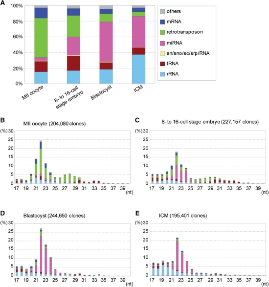
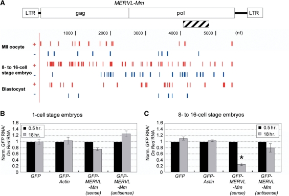
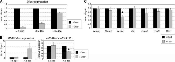
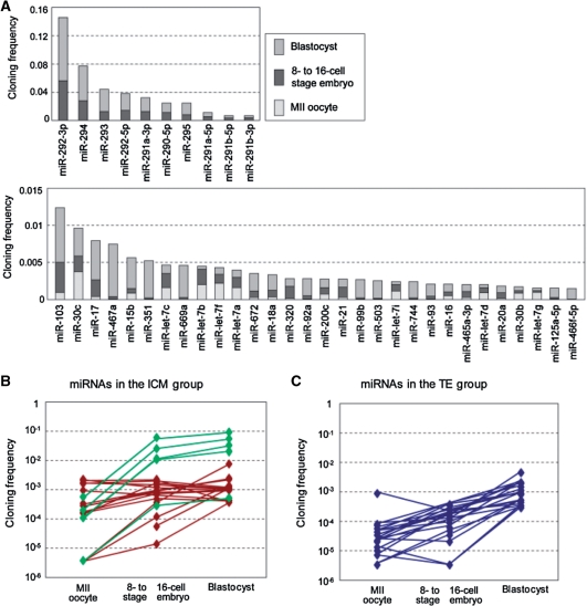
Recent studies showed that small interfering RNAs (siRNAs) and Piwi-interacting RNA (piRNA) in mammalian germ cells play important roles in retrotransposon silencing and gametogenesis. However, subsequent contribution of those small RNAs to early mammalian development remains poorly understood. We investigated the expression profiles of small RNAs in mouse metaphase II oocytes, 8-16-cell stage embryos, blastocysts and the pluripotent inner cell mass (ICM) using high-throughput pyrosequencing. Here, we show that during pre-implantation development a major small RNA class changes from retrotransposon-derived small RNAs containing siRNAs and piRNAs to zygotically synthesized microRNAs (miRNAs). Some siRNAs and piRNAs are transiently upregulated and directed against specific retrotransposon classes. We also identified miRNAs expression profiles characteristic of the ICM and trophectoderm (TE) cells. Taken together, our current study reveals a major reprogramming of functional small RNAs during early mouse development from oocyte to blastocyst.


Figures

Figure 1.
Figure 1.
Small RNA profiles in stages of early development of mouse embryos and in ICM. (A) Small RNA libraries from each stage were examined by 454 pyrosequencing followed by assignment to RNA class as described in ‘Materials and Methods’ section. The distribution of the RNA classes for each developmental stage is given as a percentage of the total annotated small RNAs. (B–E) Distribution of nucleotide lengths of annotated small RNAs as in A. The number of clones matched the mouse genome for each library is indicated in parentheses.
Figure 2.
Figure 2.
Active small RNAs derived from retrotransposons. (A) Small RNAs (19–24 nt) of the plus strand (red) and minus strand (blue) matched the MERVL-Mm retrotransposon in mouse MII oocytes and pre-implantation embryos. Hatched bar represents an insertion into the GFP 3′ UTR. (B and C) Degradation of GFP fusion mRNA carrying MERVL-Mm. The GFP, GFP-Actin, GFP-MERVL-Mm (sense), or GFP-MERVL-Mm (antisense) mRNAs was introduced together with the DsRed mRNA as a control into 1-cell and 8–16-cell stage embryos by electroporation. After 0.5 and 18 h of electroporation, total RNA was extracted from 5 to 10 embryos and the introduced mRNAs were quantified by Q-PCR. The levels of GFP and its fusion mRNAs were normalized to those of DsRed mRNAs, and then further normalized to those obtained at 0.5 h in each case [n = 3; error bars represent SEM; *P < 0.05 (t-test)].
Figure 3.
Figure 3.
Effect of Dicer-knockdown on gene expressions in pre-implantation embryo (A) Suppression of Dicer expression by siRNA. The siRNAs against Dicer (siDicer) or non-silencing control siRNAs (siCont.) were introduced into two-cell stage embryos (1.5 dpc) by electroporation, and total RNA was extracted from the electroporated embryos (2.5, 3.5 and 4.5 dpc). The expression level of Dicer was examined by Q-PCR and normalized to that of Gapdh examined as a control. The resultant level of Dicer in the presence of siDicer was further normalized to that in the presence of siControl (siCont.) as 1 (n = 3; error bars represent SEM). The data indicate that the level of Dicer mRNA in the presence of siDicer remains low until 3.5 dpc and recovers toward normal level thereafter, suggesting that Dicer-knockdown at the RNA level appears to last until 3.5 dpc. (B) MERVL-Mm and miR-99b expressions under Dicer-knockdown. The expression of MERVL-Mm, which appears to be regulated by endogenous siRNAs, and miR-99b, which appears to be a zygotic miRNA, were examined by Q-PCR using the same samples as in A. Marked increase and decrease in the levels of MERVL-Mm and miR-99b, respectively, were detected at 4.5 dpc, i.e. the effect of Dicer-knockdown on the expression of MERVL-Mm and miRNA became evident on the third day after the introduction of siDicer when the Dicer mRNA level recovered from RNAi suppression. These suggest that there is a time lag in changes in Dicer mRNA and protein levels, and we then examined the effect of Dicer-knockdown on the stability of transcripts of interest on the third day after siDicer was introduced (4.5 dpc). (C) Influence of Dicer-knockdown on gene expression. Electroporation with the siRNAs against Dicer (siDicer) and siControl (siCont.) and also preparation of total RNA were carried out as in B. The expression levels of indicated genes were examined by Q-PCR and normalized to those of Gapdh as a control. The resultant expression levels in the presence of siDicer were further normalized to those in siControl as 1. Data are averages of at least three independent experiments [error bars represent SEM; *P < 0.05 (t-test)].
Figure 4.
Figure 4.
miRNA profiles in pre-implantation mouse embryos. (A) Major miRNAs present in MII oocytes and pre-implantation embryos for the 40 most highly expressed miRNAs based on cloning frequencies. (B and C) Profiles of miRNAs abundant in ICM or TE. The 20 miRNAs in each of ICM (ICM group) (B) and TE (TE group) (C) were selected with the ratios of miRNAs (ICM/Blastocyst) (see Supplementary Table S3) and plotted for three stages from MII oocyte to blastocyst. The members belonging to the miR-290 cluster are indicated in green.
Figure 5.
Figure 5.
Proposed transition of functional small RNAs during mammalian oogenesis and early embryogenesis. Based on the present and previous study (13), possible changes in the levels of functional small RNAs from growing oocytes to pre-implantation embryos are represented schematically. Putative expression levels of zygotic siRNAs and piRNAs are represented by dotted lines and are not drawn in scale.

References

    1. Bernstein E, Caudy AA, Hammond SM, Hannon GJ. Role for a bidentate ribonuclease in the initiation step of RNA interference. Nature. 2001;409:363–366. - PubMed
    1. Bartel DP. MicroRNAs: genomics, biogenesis, mechanism, and function. Cell. 2004;116:281–297. - PubMed
    1. Aravin A, Gaidatzis D, Pfeffer S, Lagos-Quintana M, Landgraf P, Iovino N, Morris P, Brownstein MJ, Kuramochi-Miyagawa S, Nakano T, et al. A novel class of small RNAs bind to MILI protein in mouse testes. Nature. 2006;442:203–207. - PubMed
    1. Lau NC, Seto AG, Kim J, Kuramochi-Miyagawa S, Nakano T, Bartel DP, Kingston RE. Characterization of the piRNA complex from rat testes. Science. 2006;313:363–367. - PubMed
    1. Girard A, Sachidanandam R, Hannon GJ, Carmell MA. A germline-specific class of small RNAs binds mammalian Piwi proteins. Nature. 2006;442:199–202. - PubMed

Publication types