. 2010 Sep;24(9):3145-59.
doi: 10.1096/fj.09-151308. Epub 2010 Apr 12.

SIRT1 is a redox-sensitive deacetylase that is post-translationally modified by oxidants and carbonyl stress

Affiliations

SIRT1 is a redox-sensitive deacetylase that is post-translationally modified by oxidants and carbonyl stress

Samuel Caito et al. FASEB J. 2010 Sep.

Abstract

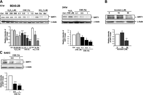
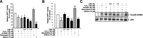
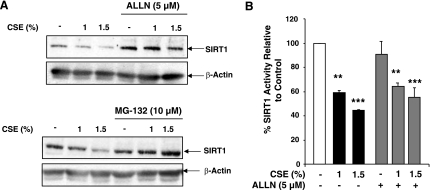
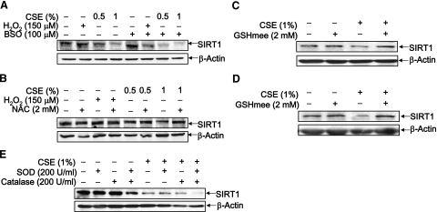
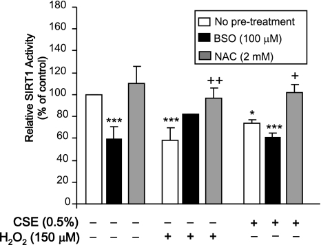
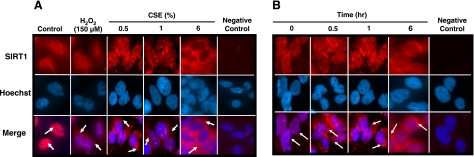
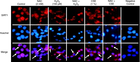
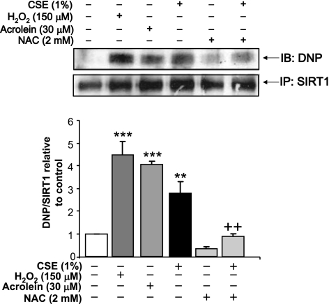
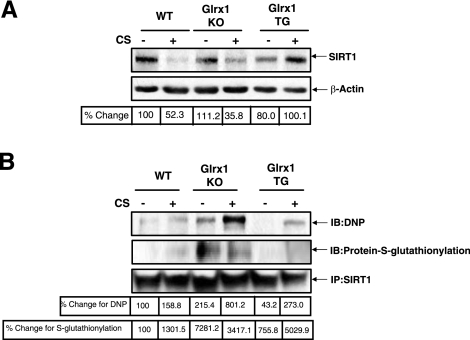
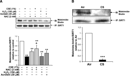
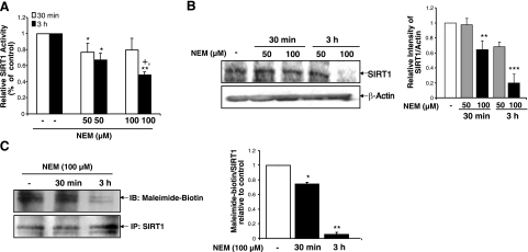
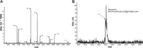
Sirtuin1 (SIRT1) deacetylase levels are decreased in chronic inflammatory conditions and aging where oxidative stress occurs. We determined the mechanism of SIRT1 redox post-translational modifications leading to its degradation. Human lung epithelial cells exposed to hydrogen peroxide (150-250 microM), aldehyde-acrolein (10-30 microM), and cigarette smoke extract (CSE; 0.1-1.5%) in the presence of intracellular glutathione-modulating agents at 1-24 h, and oxidative post-translational modifications were assayed in cells, as well as in lungs of mice lacking and overexpressing glutaredoxin-1 (Glrx1), and wild-type (WT) mice in response to cigarette smoke (CS). CSE and aldehydes dose and time dependently decreased SIRT1 protein levels, with EC(50) of 1% for CSE and 30 microM for acrolein at 6 h, and >80% inhibition at 24 h with CSE, which was regulated by modulation of intracellular thiol status of the cells. CS decreased the lung levels of SIRT1 in WT mice, which was enhanced by deficiency of Glrx1 and prevented by overexpression of Glrx1. Oxidants, aldehydes, and CS induced carbonyl modifications on SIRT1 on cysteine residues concomitant with decreased SIRT1 activity. Proteomics studies revealed alkylation of cysteine residue on SIRT1. Our data suggest that oxidants/aldehydes covalently modify SIRT1, decreasing enzymatic activity and marking the protein for proteasomal degradation, which has implications in inflammatory conditions.


Figures

Figure 1.
Figure 1.
Aldehyde and CS lower SIRT1 levels in human lung epithelial cells. A) BEAS-2B cells were treated with varying concentrations of CSE (0.1–1.5%) or H2O2 (150, 250, or 500 μM) for 6 or 24 h. Cells were also treated for 1 or 6 h with either KO2 (1 μM) or dry DMSO vehicle (Veh). Ctrl, untreated control. B) BEAS-2B cells were treated with acrolein (10 or 30 μM) for 6 h. C) SAECs were treated with CSE (0.5 and 1%) for 4 h. Whole-cell extracts from treated cells were used for immunoblot analysis for SIRT1. β-Actin was used as a housekeeping loading control. Blots are representative of ≥3 separate experiments (n=3). *P < 0.05, **P < 0.01, ***P < 0.001 vs. controls.
Figure 2.
Figure 2.
Oxidants, aldehyde, and CSE dose and time dependently decrease SIRT1 deacetylase activity. A, B) BEAS-2B cells were treated with varying concentrations of CSE (0.1, 0.5, 1, 1.5%), H2O2 (150 μM), and acrolein (30 μM) for 6 h (A) or 24 h (B). Cells were also treated with sirtinol (10 μM) and resveratrol (5 μM), a known inhibitor and activator of SIRT1, respectively. SIRT1 was immunoprecipitated from whole-cell extracts and used for the SIRT1 deacetylase activity assay. C) Cell extracts were also used for immunoblot for acetylated p53 (Lys 382) and p53 as a second measure of SIRT1 activity. Blots are representative of ≥3 separate experiments (n=3). *P < 0.05, **P < 0.01, ***P < 0.001 vs. controls.
Figure 3.
Figure 3.
SIRT1 is degraded by the proteasome in response to CSE. A) BEAS-2B cells were treated with the proteasome inhibitor N-Acetyl-Leu-Leu-Nle-CHO (ALLN; 5 μM) or MG-132 (10 μM) for 30-min pretreatment prior to a 6-h CSE (1 and 1.5%) treatment. Whole-cell extracts were analyzed by Western blot (representative of 3 separate experiments). β-Actin was used as a loading control. B) SIRT1 was immunoprecipitated from whole-cell extracts from A and used for the SIRT1 deacetylase activity assay. **P < 0.01, ***P < 0.001 vs. control.
Figure 4.
Figure 4.
Modulation of cellular thiol level regulates SIRT1 protein level. A) BEAS-2B cells were treated with BSO (100 μM) to deplete GSH levels. BSO was pretreated 18 h before a 24-h treatment with CSE (0.5 or 1%) or H2O2 (150 μM). B) BEAS-2B cells were treated with NAC (2 mM) for 2 h prior to a 24-h treatment with CSE (0.5 or 1%) or H2O2 (150 μM). C, D) BEAS-2B cells (C) and SAECs (D) were treated with glutathione monoethyl ester (GSHmee; 2 mM) for 2 h, washed off with sterile PBS, and treated with CSE (1%) for 6 h. E) PEG-conjugated SOD (200 U/ml) and catalase (200 U/ml) were given to BEAS-2B cells alone or in combination for 30 min prior to being washed off with PBS, followed by a 6-h treatment with CSE (1%). Whole-cell extracts were analyzed by immunoblot. Gels are representative of 3 separate experiments (n=3). β-Actin was used as a loading control.
Figure 5.
Figure 5.
SIRT1 activity is modulated by intracellular thiol status. BEAS-2B cells were treated with CSE (0.5%) and H2O2 (150 μM) for 24 h following an 18 h pretreatment with BSO (100 μM) or a 2 h pretreatment with NAC (2 mM). SIRT1 was immunoprecipitated from whole-cell extracts and used for the SIRT1 deacetylase assay. *P < 0.05, ***P < 0.001 vs. control; +P < 0.05, ++P < 0.01 vs. respective H2O2 or CSE treatments (n=3).
Figure 6.
Figure 6.
SIRT1 is dose- and time-dependently shuttled out of the nucleus by oxidants. A) BEAS-2B cells were treated with CSE (0.5–1.5%) or H2O2 (150 μM) for 1 h. B) BEAS-2B cells were treated with CSE (0.5%) for 30 min, 1 h, and 6 h. Cells were fixed with 4% paraformaldehyde and used for immunostaining. SIRT1 was visualized by using goat anti-rabbit secondary antibody conjugated to Alexa Fluor 594 dye. Nuclei were stained with Hoechst 33342 dye. Untreated cells that received Hoechst 33342 and secondary antibody but no SIRT1 primary antibody was used as a negative control. Arrows denote SIRT1. Images represent 3 separate experiments (n=3).
Figure 7.
Figure 7.
Nucleocytoplasmic shuttling of SIRT1 by oxidative stress is reversed by NAC. BEAS-2B cells were treated with NAC (2 mM) for 2 h prior to a 1-h treatment with either CSE (1%) or H2O2 (150 μM). Cells were fixed with 4% paraformaldehyde and used for immunostaining. SIRT1 was visualized by using goat anti-rabbit secondary antibody conjugated to Alexa Fluor 594 dye. Nuclei were stained with Hoechst 33342 dye. Untreated cells that received Hoechst 33342 and secondary antibody but no SIRT1 primary antibody was used as a negative control. Arrows denote SIRT1. Images represent 3 separate experiments (n=3).
Figure 8.
Figure 8.
Oxidative stress and CSE induce carbonylation on SIRT1. SIRT1 was immunoprecipitated (IP) from whole-cell extracts of BEAS-2B cells treated for 1 h with acrolein (30 μM), H2O2 (150 μM), and CSE (1%) in the presence or absence of a 2-h pretreatment with NAC (2 mM). Equal amount (100 μg) of immunoprecipitated SIRT1 protein was used for immunoblotting (IB). Carbonylation was detected by first derivitizing the samples with DNPH and immunoblotting with an anti-DNP antibody. Blot is representative of ≥3 separate experiments **P < 0.01, ***P < 0.001 vs. control; ++P < 0.01 vs. CSE.
Figure 9.
Figure 9.
Reversible covalent modification on SIRT1 by CS does not affect total SIRT1 levels. A) Whole-lung extracts were blotted for SIRT1 levels from WT C57BL/6J, Glrx1-KO, and Glrx1-Tg mice exposed to air or CS (300 mg/m3 TPM) for 3 d and sacrificed 24 h after last exposure, as described in Materials and Methods. B) Whole lung cell extract (100 μg) was immunoprecipitated and S-glutathionylated, and total carbonylated SIRT1 levels were detected by immunoblot with anti-GSH and anti-DNP antibodies. Blots are representative of 3 separate experiments (n=3).
Figure 10.
Figure 10.
Altering cell thiol status affects post-translational modifications on cysteine residues of SIRT1 in response to aldehydes and oxidants. SIRT1 was immunoprecipitated (IP) from whole-cell extracts of BEAS-2B cells treated for 6 h with acrolein (30 μM), H2O2 (150 μM), and CSE (1%) in the presence or absence of a 2-h pretreatment with NAC (2 mM) (A) or wild type C57BL/6J mice exposed to air or CS (B). Free cysteine residues were labeled using maleimide-PEO2-biotin and visualized using immunoblot with streptavidin conjugated to horseradish peroxidase. Blot is representative of three separate experiments (n=3). **P < 0.01, ***P < 0.001 vs. controls; +P < 0.05 vs. CSE (1%).
Figure 11.
Figure 11.
Cysteine alkylation decreases SIRT1 protein levels and enzymatic activity. A) BEAS-2B cells were treated with NEM (50 or 100 μM) for 30 min or 3 h, and SIRT1 was immunoprecipitated from whole-cell extracts for measurement of deacetylase activity. B) Extracts were also used for immunoblot for SIRT1 protein levels. β-Actin was used as a loading control. C) Nonalkylated/free cysteine residues on immunoprecipitated SIRT1 were labeled using maleimide-PEO2-biotin and visualized using immunoblot with streptavidin conjugated to horseradish peroxidase. Blots are representative of 3 separate experiments (n=3). *P < 0.05, **P < 0.01, ***P < 0.001 vs. controls; +P < 0.05 vs. NEM (50 μM) at 3 h.
Figure 12.
Figure 12.
MALDI-TOF/TOF MS spectra of SIRT1 modified by reactive aldehydes. A) Recombinant human SIRT1 (30 μg) was modified with 4-HNE (30 μM) for 18 h at 37°C and analyzed by MALDI TOF/TOF mass spectrometry (AutoflexIII TOF/TOF MALDI mass spectrometer; Bruker Daltonics). Data were analyzed using Mascot 2.1.04. B) Spectra of the sequence (residues 467–492) containing a cysteine residue at 482 modified by 4-HNE; modified cysteine is denoted by bold and underscored font.

References

    1. Elliott P J, Jirousek M. Sirtuins: novel targets for metabolic disease. Curr Opin Investig Drugs. 2008;9:371–378. - PubMed
    1. Michan S, Sinclair D. Sirtuins in mammals: insights into their biological function. Biochem J. 2007;404:1–13. - PMC - PubMed
    1. Westphal C H, Dipp M A, Guarente L. A therapeutic role for sirtuins in diseases of aging? Trends Biochem Sci. 2007;32:555–560. - PubMed
    1. Chen J, Zhou Y, Mueller-Steiner S, Chen L F, Kwon H, Yi S, Mucke L, Gan L. SIRT1 protects against microglia-dependent amyloid-beta toxicity through inhibiting NF-κB signaling. J Biol Chem. 2005;280:40364–40374. - PubMed
    1. Vogelmeier C, Bals R. Chronic obstructive pulmonary disease and premature aging. Am J Respir Crit Care Med. 2007;175:1217–1218. - PubMed

Publication types

MeSH terms