. 2010 Jun;78(6):2607-19.
doi: 10.1128/IAI.00134-10. Epub 2010 Apr 12.

Francisella tularensis DeltapyrF mutants show that replication in nonmacrophages is sufficient for pathogenesis in vivo

Affiliations

Francisella tularensis DeltapyrF mutants show that replication in nonmacrophages is sufficient for pathogenesis in vivo

Joseph Horzempa et al. Infect Immun. 2010 Jun.

Abstract

The pathogenesis of Francisella tularensis has been associated with this bacterium's ability to replicate within macrophages. F. tularensis can also invade and replicate in a variety of nonphagocytic host cells, including lung and kidney epithelial cells and hepatocytes. As uracil biosynthesis is a central metabolic pathway usually necessary for pathogens, we characterized DeltapyrF mutants of both F. tularensis LVS and Schu S4 to investigate the role of these mutants in intracellular growth. As expected, these mutant strains were deficient in de novo pyrimidine biosynthesis and were resistant to 5-fluoroorotic acid, which is converted to a toxic product by functional PyrF. The F. tularensis DeltapyrF mutants could not replicate in primary human macrophages. The inability to replicate in macrophages suggested that the F. tularensis DeltapyrF strains would be attenuated in animal infection models. Surprisingly, these mutants retained virulence during infection of chicken embryos and in the murine model of pneumonic tularemia. We hypothesized that the F. tularensis DeltapyrF strains may replicate in cells other than macrophages to account for their virulence. In support of this, F. tularensis DeltapyrF mutants replicated in HEK-293 cells and normal human fibroblasts in vitro. Moreover, immunofluorescence microscopy showed abundant staining of wild-type and mutant bacteria in nonmacrophage cells in the lungs of infected mice. These findings indicate that replication in nonmacrophages contributes to the pathogenesis of F. tularensis.


Figures

FIG. 1.
FIG. 1.
F. tularensis ΔpyrF mutants are uracil auxotrophs and resistant to FOA. LVS strains were spread onto CDM with or without uracil (left) or on CHAB with or without FOA (right). The abbreviation “comp” refers to the complementing construct, pF8pyrFfull2, and “vector” designates the empty shuttle plasmid, pFNLTP8. Similar results were observed with the cognate F. tularensis Schu S4 strains (not shown). Images shown are representative of at least four independent experiments.
FIG. 2.
FIG. 2.
Promoter characterization of F. tularensis pyrF. (A) Genomic arrangement of pyrF where the three predicted −10 and −35 promoter regions are shaded (BPROM; www.softberry.ru). The start codon of pyrF is boxed and in boldface type. (B) The indicated bacterial strains were spread onto CDM with or without uracil. The plasmid PII ppyrF refers to the construct pF8pyrFpart, and “vector” designates the empty shuttle plasmid, pFNLTP8. PII ppyrF contains sequence up to the site designated PII (A). The fully complementing vector (not shown in this figure) contains coding sequence up to the PI site. (C) Macrophages were infected with the designated strains (MOI, 500) using a gentamicin protection assay. Macrophages were lysed at the indicated time points, and lysates were diluted and plated to enumerate intracellular CFU. Data shown are means ± standard errors of the means (SEM) of results from triplicate wells within one experiment.
FIG. 3.
FIG. 3.
Heterologous expression of F. tularensis pyrF rescues the uracil auxotrophy of E. coli SKP10. E. coli SKP10 (pyrF287) was transformed with pF8pyrFfull2 (carrying pyrFLVS) or pFNLTP8 (empty vector) and was plated on LB supplemented with ampicillin (Ap) and kanamycin (Km) or on CDM. This experiment was performed on two separate occasions, with similar results.
FIG. 4.
FIG. 4.
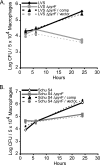
F. tularensis ΔpyrF mutants do not replicate in primary human macrophages. Macrophages were infected with the designated LVS (A) or Schu S4 (B) strains (MOI, 500) using a gentamicin protection assay. Macrophages were lysed at the indicated time points, and lysates were diluted and plated to enumerate intracellular CFU. Data shown are means ± SEM of results from triplicate wells within one experiment and are representative of three or four experiments performed using cells from separate donors. The number of viable Schu S4 ΔpyrF CFU seen after 24 h in this experiment was the most among the experiments conducted; other experiments showed a decline in Schu S4 ΔpyrF numbers similar to that observed for LVS ΔpyrF (data not shown). The abbreviation “comp” refers to the complementing construct, pF8pyrFfull2, and “vector” designates the empty shuttle plasmid, pFNLTP8. For growth in macrophages at 24 h, P was <0.001 for LVS versus LVS ΔpyrF, and P was <0.001 for Schu S4 versus Schu S4 ΔpyrF.
FIG. 5.
FIG. 5.
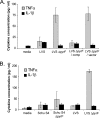
F. tularensis LVS ΔpyrF, but not Schu S4 ΔpyrF, stimulates primary macrophages to produce the proinflammatory cytokine TNF-α. Data are depicted as mean ± SEM levels of TNF-α and IL-1β secretion by primary human macrophages following a 24-h culture with the indicated strains (MOI, 10). Each panel represents a combination of three individual experiments using cells from separate blood donors. For panel B, LVS and LVS ΔpyrF were included in a single experiment for comparison. Uninfected control wells contained macrophages with no bacteria added (media). For TNF-α in panel A, P was <0.05 for LVS versus LVS ΔpyrF; for TNF-α in panel B, P was >0.05 for Schu S4 versus Schu S4 ΔpyrF.
FIG. 6.
FIG. 6.
F. tularensis ΔpyrF mutants retain virulence in animal models of infection. Chicken embryos (A) or mice (B) were infected with the indicated strains. Data represent a combination of 3 (A) or 2 (B) experiments and are shown as Kaplan-Meier survival curves depicting percent survival over time. (A) Chicken embryos were infected with ∼103, ∼105, or ∼107 CFU. All bacterial strains were used to infect chicken embryos in three experiments except LVS ΔdeoB, which was used in only one trial. Twenty embryos were used for LVS ΔpyrF, 16 for LVS ΔpyrF/pF8pyrFfull2 (comp), 16 for LVS ΔpyrF/pFNLTP8 (vector), and 6 for LVS ΔdeoB. For each experiment, the actual numbers of CFU administered to the chicken embryos were as follows: for LVS, 8.0 × 10², 2.8 × 10⁵, and 5.0 × 10⁷; for LVS ΔpyrF, 1.6 × 10³, 9.0 × 10⁴, and 1.2 × 10⁷; for LVS ΔpyrF/comp, 1.1 × 10³, 1.8 × 10⁵, and 4.6 × 10⁷; for ΔpyrF/vector, 6.2 × 10², 2.6 × 10⁵, and 1.7 × 10⁷; and for LVS ΔdeoB, 2.5 × 10². P was <0.0001 for LVS versus LVS ΔdeoB. (B) Mice were infected (i.t.) with ∼101 or ∼103 CFU of each strain. Ten mice (two experiments) were used for each infecting strain except LVS, for which five mice (included in only one experiment) were used. For each experiment, the actual numbers of CFU administered to mice were as follows: for Schu S4, 37 and 350; for Schu S4 ΔpyrF, 14 and 500; for Schu S4 ΔpyrF/comp, 19 and 250; for Schu S4 ΔpyrF/vector, 49 and 850; and for LVS, 850. A subset of mice was sacrificed at 2 h postinfection, and their lungs were homogenized and plated to confirm the efficacy of bacterial delivery to pulmonary tissue, which indicated comparable rates of delivery for all strains (not shown). P was 0.0002 for LVS versus Schu S4 ΔpyrF; P was <0.0001 for Schu S4 versus Schu S4 ΔpyrF.
FIG. 7.
FIG. 7.
F. tularensis Schu S4 ΔpyrF exhibits dissemination to peripheral organs in a murine respiratory tularemia model. Mice were infected (i.t.) with ∼102 CFU of F. tularensis Schu S4, Schu S4 ΔpyrF, or LVS. The average numbers of CFU administered to mice over both experiments were as follows: for Schu S4, 200; for Schu S4 ΔpyrF, 190; and for LVS, 140. A subset of mice was sacrificed at 2 h postinfection, and their lungs were homogenized and plated to confirm the efficacy of bacterial delivery to pulmonary tissue, which indicated comparable rates of delivery for all strains (not shown). At day 2 or 4 postinfection, mice were sacrificed and the designated organs and tissues were harvested, homogenized, diluted, and plated for CFU enumeration. The limit of detection was 100 CFU per organ (or per ml blood), except the liver, which was 200 CFU. Data, displayed as mean numbers of CFU ± SEM, represent a combination of two independent experiments, each having three mice per bacterial strain per time point. The comparisons in which P was <0.05 are designated by brackets.
FIG. 8.
FIG. 8.
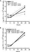
F. tularensis ΔpyrF mutants retain the ability to replicate in nonmacrophages. HEK-293 cells were infected with the designated LVS (A) or Schu S4 (B) strains (MOI, 500) using a gentamicin protection assay. HEK-293 cells were lysed at the indicated time points, and the lysates were diluted and plated to enumerate intracellular CFU. Data shown are means ± SEM of results from triplicate wells within one experiment and are representative of at least three independent experiments. The abbreviations are the same as those in Fig. 4. For growth in HEK-293 cells at 24 h, P was 0.0083 for LVS versus LVS ΔpyrF, and P was 0.076 for Schu S4 versus Schu S4 ΔpyrF.
FIG. 9.
FIG. 9.
F. tularensis ΔpyrF mutants retain the ability to replicate in normal fibroblasts. NHDF cells were infected with the designated LVS (A) or Schu S4 (B) strains (MOI, 500) using a gentamicin protection assay. NHDF cells were lysed at the indicated times, and the lysates were diluted and plated to enumerate CFU. Data shown are means ± SEM of results from triplicate wells within one experiment and are representative of at least three independent experiments. The abbreviations are the same as those in Fig. 4.
FIG. 10.
FIG. 10.
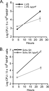
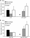
Intracellular growth rate of wild-type F. tularensis and ΔpyrF mutants. Mean generation time (± SEM) was calculated for bacterial replication in primary human macrophages, HEK-293 cells, or NHDF cells from at least three combined independent experiments. Panel A shows the mean generation time comparison of LVS and LVS ΔpyrF, and panel B depicts the Schu S4 and Schu S4 ΔpyrF data. For both panels, the generation time of the ΔpyrF strains in macrophages could not be calculated, because the mean numbers of viable CFU across experiments were lower at 24 h than at 2 h postinfection (asterisk).
FIG. 11.
FIG. 11.
Immunofluorescence microscopy of lung sections from mice infected with F. tularensis Schu S4 or Schu S4 ΔpyrF. Mice were infected i.t. as described in the legend to Fig. 7. On day 4 postinfection, lungs were harvested from mice infected with Schu S4 (A) or Schu S4 ΔpyrF (B), were fixed in formalin, and were subsequently embedded in paraffin. Deparaffinized lung sections were probed with rat anti-mouse F4/80 to detect macrophages and with rabbit anti-F. tularensis. The secondary antibodies used were Alexa Fluor 555 goat anti-rat immunoglobulin and Alexa Fluor 488 donkey anti-rabbit immunoglobulin. DNA was stained with DAPI. For both panels, the color images represent a three-color merge (red, F4/80; green, F. tularensis; and blue, DAPI). The first color image represents low-power magnification, and the second image is a high-power magnification of the boxed section. In the low-power images, the asterisk designates a major airway. The scale bars in the upper right hand corners of the images in panel A represent a length of 20 μm.
FIG. 12.
FIG. 12.
Quantification of Schu S4 and ΔpyrF mutant staining during pulmonary infection. Lung sections from mice infected with Schu S4 and Schu S4 ΔpyrF were harvested at day 2 or 4 postinfection. F. tularensis fluorescence (green) was quantified from infected F4/80+ and F4/80 cells using MicroSuite Basic Edition software. Data shown are from two independent infections and comprise at least three mice per time point. Fluorescence intensity was measured from regions exhibiting F. tularensis colonization in which three separate fields per section were quantified from each tissue section. (A) Cumulative green fluorescence intensity in F4/80+ and F4/80 cells. The average cumulative green fluorescence intensities ± standard errors (SE) were as follows: for Schu S4 F4/80+, 50.1 ± 16.0 (day 2) and 72.0 ± 12.0 (day 4); for Schu S4 F4/80, 120.0 ± 48.0 (day 2) and 89.6 ± 42.3 (day 4); for Schu S4 ΔpyrF F4/80+, 20.0 ± 8.8 (day 2) and 27.4 ± 6.7 (day 4); and for Schu S4 ΔpyrF F4/80, 62.0 ± 23.3 (day 2) and 88.3 ± 12.6 (day 4). The ratio of F. tularensis fluorescence in F4/80+ cells to the total (F4/80+ plus F4/80) was significantly greater in wild-type Schu S4 than in the ΔpyrF mutant (χ2 = 4.637; P < 0.05). (B) Average green fluorescence per μm2 host cell. The percentage of green fluorescence intensity per μm2 within F4/80+ host cells was significantly greater for Schu S4 (expected) than for Schu S4 ΔpyrF (observed) (χ2 = 12.96; P < 0.001).

Similar articles

Cited by

References

    1. Abril, C., H. Nimmervoll, P. Pilo, I. Brodard, B. Korczak, S. Markus, R. Miserez, and J. Frey. 2008. Rapid diagnosis and quantification of Francisella tularensis in organs of naturally infected common squirrel monkeys (Saimiri sciureus). Vet. Microbiol. 127:203-208. - PubMed
    1. Bach, M. L., F. Lacroute, and D. Botstein. 1979. Evidence for transcriptional regulation of orotidine-5′-phosphate decarboxylase in yeast by hybridization of mRNA to the yeast structural gene cloned in Escherichia coli. Proc. Natl. Acad. Sci. U. S. A. 76:386-390. - PMC - PubMed
    1. Barker, J. R., A. Chong, T. D. Wehrly, J. J. Yu, S. A. Rodriguez, J. Liu, J. Celli, B. P. Arulanandam, and K. E. Klose. 2009. The Francisella tularensis pathogenicity island encodes a secretion system that is required for phagosome escape and virulence. Mol. Microbiol. 74:1459-1470. - PMC - PubMed
    1. Barker, J. R., and K. E. Klose. 2007. Molecular and genetic basis of pathogenesis in Francisella tularensis. Ann. N. Y. Acad. Sci. 1105:138-159. - PubMed
    1. Boeke, J. D., F. LaCroute, and G. R. Fink. 1984. A positive selection for mutants lacking orotidine-5′-phosphate decarboxylase activity in yeast: 5-fluoro-orotic acid resistance. Mol. Gen. Genet. 197:345-346. - PubMed

Publication types

MeSH terms