. 2010 Jun;78(6):2631-43.
doi: 10.1128/IAI.00173-10. Epub 2010 Apr 12.

Characterization and serologic analysis of the Treponema pallidum proteome

Affiliations

Characterization and serologic analysis of the Treponema pallidum proteome

Melanie A McGill et al. Infect Immun. 2010 Jun.

Abstract

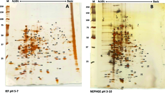
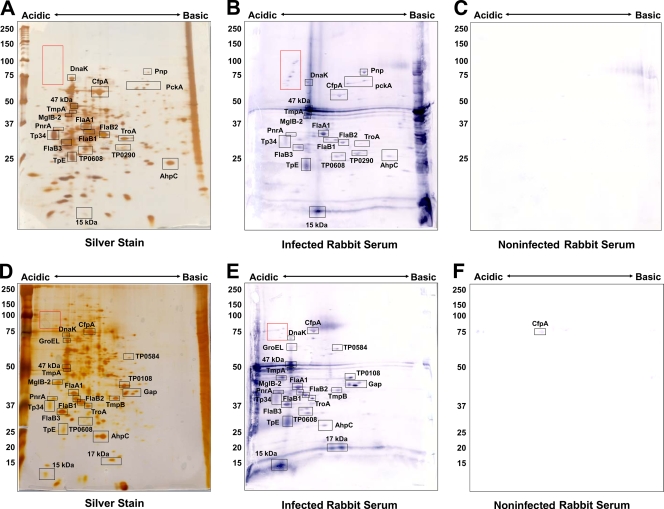
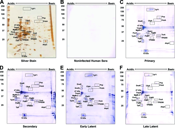
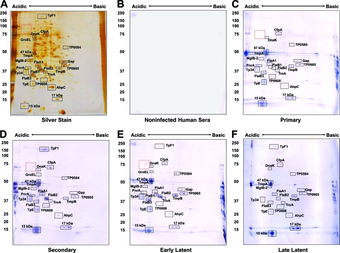
Treponema pallidum subsp. pallidum is the causative agent of syphilis, a sexually transmitted disease characterized by widespread tissue dissemination and chronic infection. In this study, we analyzed the proteome of T. pallidum by the isoelectric focusing (IEF) and nonequilibrating pH gel electrophoresis (NEPHGE) forms of two-dimensional gel electrophoresis (2DGE), coupled with matrix-assisted laser desorption ionization-time of flight (MALDI-TOF) analysis. We determined the identity of 148 T. pallidum protein spots, representing 88 T. pallidum polypeptides; 63 of these polypeptides had not been identified previously at the protein level. To examine which of these proteins are important in the antibody response to syphilis, we performed immunoblot analysis using infected rabbit sera or human sera from patients at different stages of syphilis infection. Twenty-nine previously described antigens (predominantly lipoproteins) were detected, as were a number of previously unidentified antigens. The reactivity patterns obtained with sera from infected rabbits and humans were similar; these patterns included a subset of antigens reactive with all serum samples tested, including CfpA, MglB-2, TmpA, TmpB, flagellins, and the 47-kDa, 17-kDa, and 15-kDa lipoproteins. A unique group of antigens specifically reactive with infected human serum was also identified and included the previously described antigen TpF1 and the hypothetical proteins TP0584, TP0608, and TP0965. This combined proteomic and serologic analysis further delineates the antigens potentially useful as vaccine candidates or diagnostic markers and may provide insight into the host-pathogen interactions that occur during T. pallidum infection.


Figures

FIG. 1.
FIG. 1.
Two-dimensional gel electrophoresis of T. pallidum proteins. T. pallidum lysates were separated by IEF at pH 5 to 7 (A) or NEPHGE at pH 3.5 to 10 (B) in the first dimension, followed by 8 to 20% SDS-PAGE in the second dimension. Gels were subsequently silver stained for protein visualization. Acidic and basic ends are denoted, and relative molecular mass markers (in kilodaltons) are indicated to the left of each gel. A T. pallidum lysate, resolved in the second dimension only, is shown at the right side of the IEF pH 5-to-7 gel (A). The identities of the numbered spots are presented in Table 1. Arrows indicate spots that were submitted separately for MALDI-TOF MS but returned the same identity. Circles demarcate some closely spaced spots to indicate more clearly which spots are labeled.
FIG. 2.
FIG. 2.
Correlation between predicted molecular weights of T. pallidum proteins and Mr values obtained in 2DGE patterns in this study. Molecular weights take into account removal of predicted signal peptides. Apparent degradation products were excluded.
FIG. 3.
FIG. 3.
Immunoreactivity of T. pallidum proteins separated by 2DGE with rabbit sera. T. pallidum lysates were separated by IEF at pH 5 to 7 (A to C) or NEPHGE at pH 3.5 to 10 (D to F) in the first dimension, followed by 8 to 20% SDS-PAGE in the second dimension. Gels were subsequently silver stained (A, D) or immunoblotted with a 1:1,000 dilution of infected (B, E) or noninfected (C, F) rabbit sera. Black boxed areas indicate major polypeptides that were reactive with each serum pool. Red boxed areas indicate unidentified acidic proteins. Acidic and basic ends are denoted, and relative molecular mass markers (in kilodaltons) are indicated to the left of each gel.
FIG. 4.
FIG. 4.
Immunoreactivity of T. pallidum proteins separated by IEF (pH 5 to 7) 2DGE with human sera. T. pallidum lysates were separated by IEF at pH 5 to 7 in the first dimension, followed by 8 to 20% SDS-PAGE in the second dimension. Gels were subsequently silver stained (A) or immunoblotted with a 1:500 dilution of pooled human sera from healthy blood donors (B), primary-syphilis patients (C), secondary-syphilis patients (D), early-latent-syphilis patients (E) or late-latent-syphilis patients (F). Black boxed areas indicate major polypeptides that were reactive with each serum pool. Acidic and basic ends are denoted, and relative molecular mass markers (in kilodaltons) are indicated to the left of each gel.
FIG. 5.
FIG. 5.
Immunoreactivity of T. pallidum proteins separated by NEPHGE (pH 3.5 to 10) 2DGE with human sera. T. pallidum lysates were separated by NEPHGE at pH 3.5 to 10 in the first dimension, followed by 8 to 20% SDS-PAGE in the second dimension. Gels were subsequently silver stained (A) or immunoblotted with a 1:500 dilution of pooled human sera from healthy blood donors (B), primary-syphilis patients (C), secondary-syphilis patients (D), early-latent-syphilis patients (E), or late-latent-syphilis patients (F). Black boxed areas indicate major polypeptides that were reactive with each serum pool. Acidic and basic ends are denoted, and relative molecular mass markers (in kilodaltons) are indicated to the left of each gel.

Similar articles

Cited by

References

    1. Akins, D. R., B. K. Purcell, M. M. Mitra, M. V. Norgard, and J. D. Radolf. 1993. Lipid modification of the 17-kilodalton membrane immunogen of Treponema pallidum determines macrophage activation as well as amphiphilicity. Infect. Immun. 61:1202-1210. - PMC - PubMed
    1. Borenstein, L. A., J. D. Radolf, T. E. Fehniger, D. R. Blanco, J. N. Miller, and M. A. Lovett. 1988. Immunization of rabbits with recombinant Treponema pallidum surface antigen 4D alters the course of experimental syphilis. J. Immunol. 140:2415-2421. - PubMed
    1. Brinkman, M. B., M. McKevitt, M. McLoughlin, C. Perez, J. Howell, G. M. Weinstock, S. J. Norris, and T. Palzkill. 2006. Reactivity of antibodies from syphilis patients to a protein array representing the Treponema pallidum proteome. J. Clin. Microbiol. 44:888-891. - PMC - PubMed
    1. Centers for Disease Control and Prevention. 2006. Together we can. The National Plan to Eliminate Syphilis from the United States. U.S. Department of Health and Human Services, Washington, DC.
    1. Cox, D. L., P. Chang, A. W. McDowall, and J. D. Radolf. 1992. The outer membrane, not a coat of host proteins, limits antigenicity of virulent Treponema pallidum. Infect. Immun. 60:1076-1083. - PMC - PubMed

Publication types

MeSH terms

LinkOut - more resources