. 2010 Apr 27;107(17):7799-804.
doi: 10.1073/pnas.1003586107. Epub 2010 Apr 12.

Fetal liver hepatic progenitors are supportive stromal cells for hematopoietic stem cells

Affiliations

Fetal liver hepatic progenitors are supportive stromal cells for hematopoietic stem cells

Song Chou et al. Proc Natl Acad Sci U S A. .

Abstract

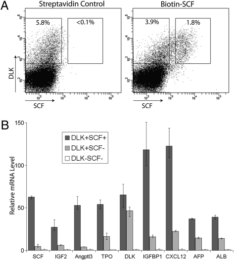
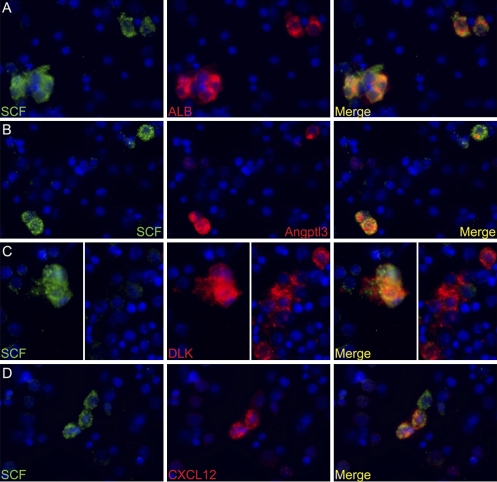
Previously we showed that the ~2% of fetal liver cells reactive with an anti-CD3epsilon monoclonal antibody support ex vivo expansion of both fetal liver and bone marrow hematopoietic stem cells (HSCs); these cells express two proteins important for HSC ex vivo expansion, IGF2, and angiopoietin-like 3. Here we show that these cells do not express any CD3 protein and are not T cells; rather, we purified these HSC-supportive stromal cells based on the surface phenotype of SCF(+)DLK(+). Competitive repopulating experiments show that SCF(+)DLK(+) cells support the maintenance of HSCs in ex vivo culture. These are the principal fetal liver cells that express not only angiopoietin-like 3 and IGF2, but also SCF and thrombopoietin, two other growth factors important for HSC expansion. They are also the principal fetal liver cells that express CXCL12, a factor required for HSC homing, and also alpha-fetoprotein (AFP), indicating that they are fetal hepatic stem or progenitor cells. Immunocytochemistry shows that >93% of the SCF(+) cells express DLK and Angptl3, and a portion of SCF(+) cells also expresses CXCL12. Thus SCF(+)DLK(+) cells are a highly homogenous population that express a complete set of factors for HSC expansion and are likely the primary stromal cells that support HSC expansion in the fetal liver.


Conflict of interest statement

The authors declare no conflict of interest.

Figures

Fig. 1.
Fig. 1.
Potential stromal cells for HSC expansion in E15.5 fetal liver can be purified by SCF+DLK+ surface phenotype. (A) FACS analysis of total fetal liver cells stained with biotin-SCF and DLK antibodies. Approximately 1.8% fetal liver cells are SCF+, most of which are also DLK+. Approximately 3.9% of fetal liver cells are DLK+SCF. (B) Relative expression of mRNAs encoding HSC expansion factors IGF2, Angptl3, TPO, IGFBP1, and CXCL12 and hepatocyte markers AFP and ALB. SCF+DLK+, SCFDLK+, and SCFDLK cells were sorted by flow cytometry. The mRNA level of each gene was determined by qPCR and normalized against ribosomal RNA. The relative expression of each gene in each population was calculated, setting the level of each mRNA in SCFDLK cells as 1 (SCF+ cells were gated more stringently in this analysis than in Fig. S2A). Numbers are average of several individual repeats (n = 3); error bars indicate maximal and minimal values scored.
Fig. 2.
Fig. 2.
Coculture of purified fetal liver HSCs with fetal liver E15.5 SCF+DLK+ cells. A total of 25 sorted E15.5 fetal liver HSCs (CD45.2) were cultured in a serum-containing medium supplemented with SCF, IL6, and FLT3 for 4 days either without (B) or with (C) 2,000 sorted E15.5 SCF+DLK+ cells. A total of 25 sorted HSCs were also mixed with 2,000 sorted fetal liver E15.5 SCF+DLK+ cells and were transplanted directly into CD45.1 mice as a control (A). Each filled triangle in the figure represents the percentage repopulation of one recipient mouse, which is calculated as percentage of CD45.2+ cells divided by total number of CD45.1+ plus CD45.2+ cells in the peripheral blood. Average percentage of reconstitution for each experiment is also indicated (n = 7–9).
Fig. 3.
Fig. 3.
Expression of HSC expansion factors during liver development. Total RNAs were extracted from fetal liver E13.5–16.5 cells, neonatal liver (D3), and adult liver. The mRNA level of each gene was normalized to 18S RNA and the expression level of each gene at E13.5 was set as 1 (n = 3).
Fig. 4.
Fig. 4.
SCF+ cells in E15.5 fetal liver are also positive for ALB, Angptl3, and DLK expression but are heterogeneous for CXCL12 expression. (A) Double immunocytochemistry for SCF (green) and ALB (red) expression in total fetal liver cells. DAPI was used to stain nuclei (blue). SCF+ cells are also positive for ALB expression. (B) Double staining for SCF and Angptl3. Three SCF+ cells are shown, all of which are positive for Angptl3 expression. (C) Double staining for SCF and DLK. (Left) Cluster of SCF+ cells that are also DLK+. DLK is also expressed by other cells, as indicated (Right) by a group of DLK+ cells that are SCF. (D) Only a fraction of SCF+ cells are also CXCL12+. Shown are three SCF+ cells, two of which are CXCL12+ and one is CXCL12 (white arrow).
Fig. 5.
Fig. 5.
AFP+ fetal hepatobasts are enriched in stromal cells that express seven growth factors that support HSC maintenance, expansion, or homing. (A) FACS analysis of E15.5 fetal liver cells from Tg(AFP-GFP) mice stained by an SCF antibody as in Fig. 1; AFP+ cells are detected by GFP expression. (B) Relative expression of mRNAs encoding HSC expansion factors IGF2, Angptl3, TPO, IGFBP1, and CXCL12 and hepatocyte markers AFP and ALB in GFP+ and GFP cells sorted by flow cytometry. The mRNA level of each gene was determined by qPCR and normalized against ribosomal RNA. The relative expression of each gene in each population was calculated setting the level of each mRNA in GFP cells as in Fig. 1 (n = 3).

References

    1. Martinez-Agosto JA, Mikkola HK, Hartenstein V, Banerjee U. The hematopoietic stem cell and its niche: A comparative view. Genes Dev. 2007;21:3044–3060. - PubMed
    1. Wilson A, Trumpp A. Bone-marrow haematopoietic-stem-cell niches. Nat Rev Immunol. 2006;6:93–106. - PubMed
    1. Taichman RS. Blood and bone: Two tissues whose fates are intertwined to create the hematopoietic stem-cell niche. Blood. 2005;105:2631–2639. - PubMed
    1. Calvi LM, et al. Osteoblastic cells regulate the haematopoietic stem cell niche. Nature. 2003;425:841–846. - PubMed
    1. Kiel MJ, et al. SLAM family receptors distinguish hematopoietic stem and progenitor cells and reveal endothelial niches for stem cells. Cell. 2005;121:1109–1121. - PubMed

Publication types