. 2010 Apr 27;107(17):7999-8004.
doi: 10.1073/pnas.1003655107. Epub 2010 Apr 12.

RGS4 is a negative regulator of insulin release from pancreatic beta-cells in vitro and in vivo

Affiliations

RGS4 is a negative regulator of insulin release from pancreatic beta-cells in vitro and in vivo

Inigo Ruiz de Azua et al. Proc Natl Acad Sci U S A. .

Abstract

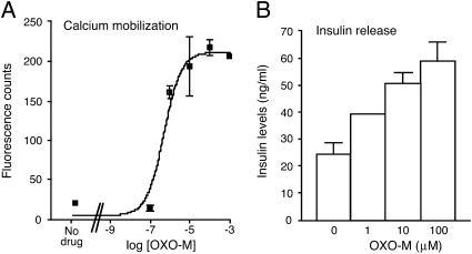
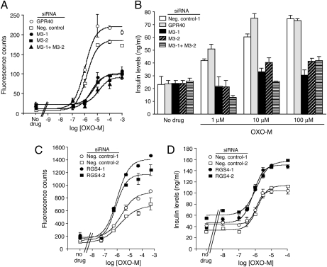
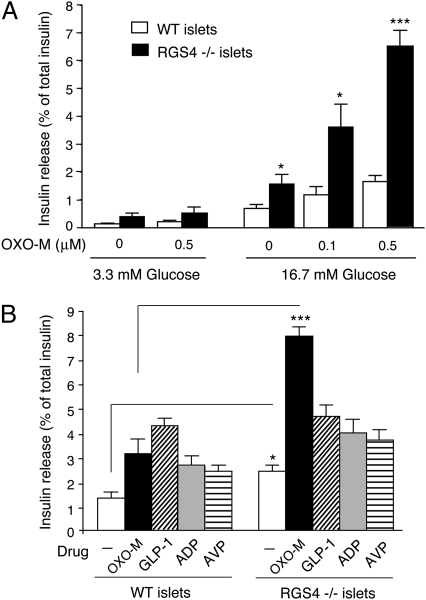
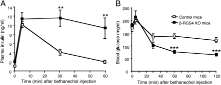
Therapeutic strategies that augment insulin release from pancreatic beta-cells are considered beneficial in the treatment of type 2 diabetes. We previously demonstrated that activation of beta-cell M(3) muscarinic receptors (M3Rs) greatly promotes glucose-stimulated insulin secretion (GSIS), suggesting that strategies aimed at enhancing signaling through beta-cell M3Rs may become therapeutically useful. M3R activation leads to the stimulation of G proteins of the G(q) family, which are under the inhibitory control of proteins known as regulators of G protein signaling (RGS proteins). At present, it remains unknown whether RGS proteins play a role in regulating insulin release. To address this issue, we initially demonstrated that MIN6 insulinoma cells express functional M3Rs and that RGS4 was by far the most abundant RGS protein expressed by these cells. Strikingly, siRNA-mediated knockdown of RGS4 expression in MIN6 cells greatly enhanced M3R-mediated augmentation of GSIS and calcium release. We obtained similar findings using pancreatic islets prepared from RGS4-deficient mice. Interestingly, RGS4 deficiency had little effect on insulin release caused by activation of other beta-cell GPCRs. Finally, treatment of mutant mice selectively lacking RGS4 in pancreatic beta-cells with a muscarinic agonist (bethanechol) led to significantly increased plasma insulin and reduced blood glucose levels, as compared to control littermates. Studies with beta-cell-specific M3R knockout mice showed that these responses were mediated by beta-cell M3Rs. These findings indicate that RGS4 is a potent negative regulator of M3R function in pancreatic beta-cells, suggesting that RGS4 may represent a potential target to promote insulin release for therapeutic purposes.


Conflict of interest statement

The authors declare no conflict of interest.

Figures

Fig. 1.
Fig. 1.
OXO-M-mediated increases in [Ca2+]i and insulin release in MIN6 cells. (A) Calcium measurements. OXO-M-induced increases in [Ca2+]i were measured in MIN6 cells grown in 96-well plates by using FLIPR technology. (B) Insulin release measurements. OXO-M–mediated increases in insulin secretion were determined in the presence of 16.7 mM glucose. Representative concentration-response curves are shown. Data points represent means ± SEM of assays carried out in triplicate. Nine additional experiments gave similar results.
Fig. 2.
Fig. 2.
Effect of siRNA-mediated M3R or RGS4 knockdown in MIN6 cells on OXO-M-induced calcium and insulin responses. Following treatment of MIN6 cells with the indicated siRNAs, OXO-M-induced increases in [Ca2+]i and insulin secretion were determined. (A–D) Effect of M3R knockdown (A and B) or RGS4 knockdown (C and D) on OXO-M-induced calcium and insulin responses. Assays were carried out with MIN6 cells that had been electroporated with the indicated siRNAs. Negative control siRNAs represent scrambled siRNAs. In the M3R knockdown assays, GPR40 siRNA was included as an additional negative control (see main text for details). OXO-M-induced increases in [Ca2+]i were measured by using FLIPR technology. OXO-M-stimulated enhancement of insulin secretion was assessed in the presence of 16.7 mM glucose. Representative concentration-response curves are shown. Data points represent means ± SEM of assays carried out in triplicate. At least three additional experiments gave similar results.
Fig. 3.
Fig. 3.
Coimmunoprecipitation of RGS4 protein and the M3R in cotransfected COS-7 cells. COS-7 cells were transfected with plasmids coding for human RGS4 and an HA-epitope-tagged version of the human M3R, either in combination (lane 1) or individually (lanes 3 and 4). For control purposes, cell membranes prepared from COS-cells expressing either RGS4 or M3R (lane 2) were mixed and processed in the same fashion as all other samples. Forty-eight hours after transfection, membrane extracts were prepared (approximately 1 mg of total protein) and subjected to immunoprecipitation (IP) using an agarose-conjugated anti-HA monoclonal antibody. RGS4 was detected via Western blotting analysis using a rabbit polyclonal antibody raised against human RGS4 (size of the immunoreactive band: approximately 24 kDa). Please note that RGS4 was only detectable in samples derived from COS-7 cells that had been cotransfected with the RGS4 and M3R plasmids. Two additional experiments gave similar results. “Input” represents cell lysates used for IP studies.
Fig. 4.
Fig. 4.
Selective augmentation of OXO-M-induced insulin release in pancreatic islets prepared from RGS4-deficient mice. (A) Concentration-dependent enhancement of OXO-M-induced insulin release in islets prepared from RGS4 KO mice (RGS4−/− mice). Isolated pancreatic islets prepared from WT and RGS4 KO mice were incubated for 1 h at 37 °C in Krebs solution containing the indicated glucose concentrations, either in the absence or the presence of OXO-M (0.1 and 0.5 μM). (B) Selective augmentation of insulin secretion from RGS4-deficient pancreatic islets by OXO-M. Isolated pancreatic islets prepared from WT and RGS4−/− mice were incubated for 1 h at 37 °C in Krebs solution containing 16.7 mM glucose. In addition, the incubation medium contained one of the following four ligands known to promote insulin secretion via activation of specific β-cell GPCRs: OXO-M (0.5 μM), GLP-1 (10 nM), ADP (100 μM), or AVP (100 nM). The amount of insulin secreted into the medium during the 1-h incubation period was normalized to the total insulin content of each well (islets plus medium). Data are expressed as means ± SEM of three independent experiments, each carried out in triplicate. *P < 0.05, ***P < 0.001, as compared to the corresponding WT value.
Fig. 5.
Fig. 5.
Enhanced insulin secretion and reduced blood glucose levels following bethanechol treatment of β-RGS4-KO mice in vivo. (A and B) β-RGS4-KO mice and control littermates received a single dose of bethanechol (2 μg/g, s.c.). Plasma insulin (A) and blood glucose (B) levels were measured at the indicated time points. Values are given as means ± SEM (n = 6 per group; freely fed 9-month-old males). **P < 0.01, ***P < 0.001, as compared to the corresponding values obtained with control mice.

References

    1. Kahn SE. The relative contributions of insulin resistance and beta-cell dysfunction to the pathophysiology of Type 2 diabetes. Diabetologia. 2003;46:3–19. - PubMed
    1. Moller DE. New drug targets for type 2 diabetes and the metabolic syndrome. Nature. 2001;414:821–827. - PubMed
    1. Regard JB, et al. Probing cell type-specific functions of Gi in vivo identifies GPCR regulators of insulin secretion. J Clin Invest. 2007;117:4034–4043. - PMC - PubMed
    1. Ahrén B. Islet G protein-coupled receptors as potential targets for treatment of type 2 diabetes. Nat Rev Drug Discov. 2009;8:369–385. - PubMed
    1. Pierce KL, Premont RT, Lefkowitz RJ. Seven-transmembrane receptors. Nat Rev Mol Cell Biol. 2002;3:639–650. - PubMed

Publication types