. 2010 Apr 27;107(17):7875-80.
doi: 10.1073/pnas.1003345107. Epub 2010 Apr 12.

Tumor-infiltrating NY-ESO-1-specific CD8+ T cells are negatively regulated by LAG-3 and PD-1 in human ovarian cancer

Affiliations

Tumor-infiltrating NY-ESO-1-specific CD8+ T cells are negatively regulated by LAG-3 and PD-1 in human ovarian cancer

Junko Matsuzaki et al. Proc Natl Acad Sci U S A. .

Abstract

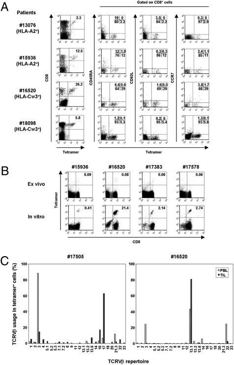
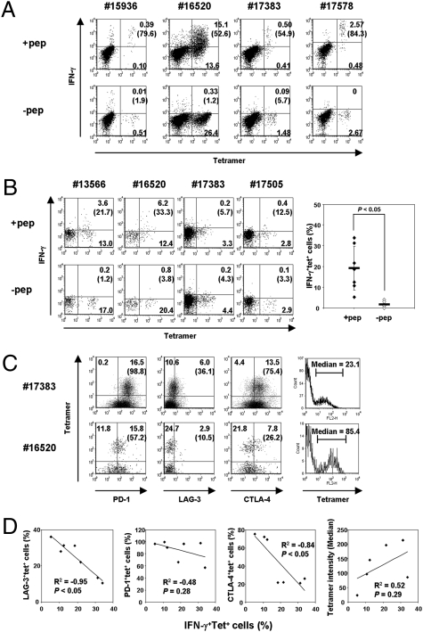
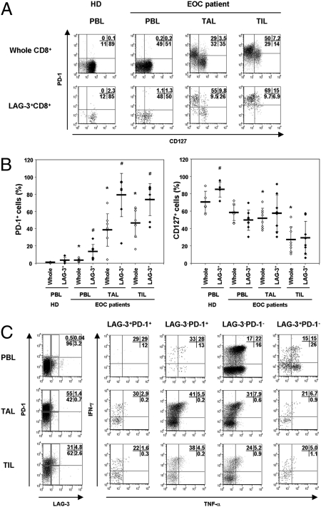
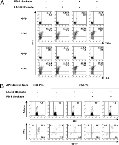
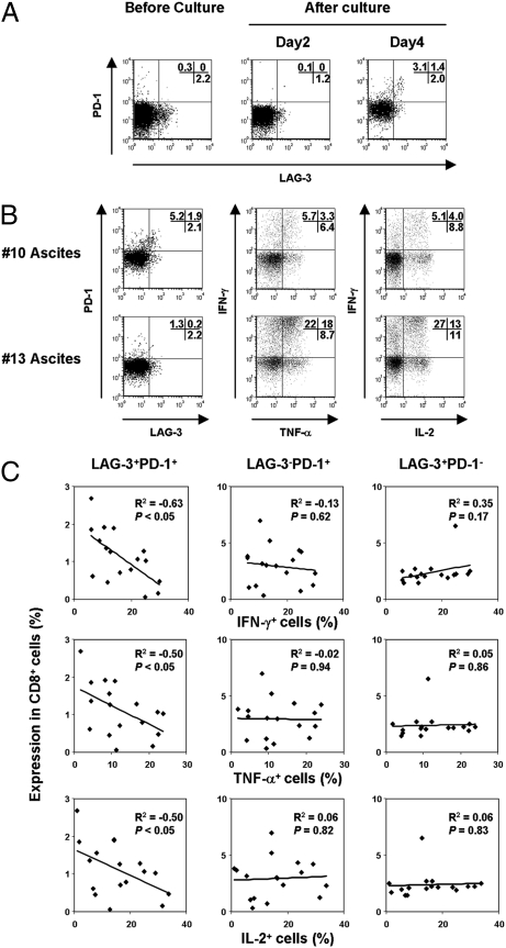
NY-ESO-1 is a "cancer-testis" antigen frequently expressed in epithelial ovarian cancer (EOC) and is among the most immunogenic tumor antigens defined to date. In an effort to understand in vivo tolerance mechanisms, we assessed the phenotype and function of NY-ESO-1-specific CD8(+) T cells derived from peripheral blood lymphocytes (PBLs), tumor-infiltrating lymphocytes (TILs), and tumor-associated lymphocytes (TALs) of EOC patients with NY-ESO-1-expressing tumors, with or without humoral immunity to NY-ESO-1. Whereas NY-ESO-1-specific CD8(+) T cells were readily detectable ex vivo with tetramers in TILs and TALs of seropositive patients, they were only detectable in PBLs following in vitro stimulation. Compared with PBLs, tumor-derived NY-ESO-1-specific CD8(+) T cells demonstrated impaired effector function, preferential usage of dominant T-cell receptor, and enriched coexpression of inhibitory molecules LAG-3 and PD-1. Expression of LAG-3 and PD-1 on CD8(+) T cells was up-regulated by IL-10, IL-6 (cytokines found in tumor ascites), and tumor-derived antigen-presenting cells. Functionally, CD8(+)LAG-3(+)PD-1(+) T cells were more impaired in IFN-gamma/TNF-alpha production compared with LAG-3(+)PD-1(-) or LAG-3(-)PD-1(-) subsets. Dual blockade of LAG-3 and PD-1 during T-cell priming efficiently augmented proliferation and cytokine production by NY-ESO-1-specific CD8(+) T cells, indicating that antitumor function of NY-ESO-1-specific CD8(+) T cells could potentially be improved by therapeutic targeting of these inhibitory receptors.


Conflict of interest statement

The authors declare no conflict of interest.

Figures

Fig. 1.
Fig. 1.
NY-ESO-1–specific CD8+ cells at the tumor site. (A) Ex vivo phenotypic analysis of NY-ESO-1 tetramer+ cells in TILs and TALs of NY-ESO-1-seropositive EOC patients. The four examples shown are representative of 15 individual samples. The HLA tetramers for individual patients are in parentheses. (B) The frequency of tetramer+ cells in PBLs before and after in vitro peptide presensitization. The number in the right upper quadrant indicates percentage of tetramer+CD8+ cells. (C) Comparison of TCR Vβ use of HLA-Cw*03-ESO92–100 tetramer+ cells derived from PBLs and TILs.
Fig. 2.
Fig. 2.
Impaired effector function of CD8+ T cells at the ovarian tumor site correlates with the expression of coinhibitory molecules. (A and B) NY-ESO-1–specific IFN-γ production from PBLs following in vitro presensitization (A). Ex vivo IFN-γ production from TILs or TALs. The graph indicates the average and statistics of peptide-specific IFN-γ production from seven samples (B). The number in parentheses indicates percentage of IFN-γ-producing cells among NY-ESO-1–specific tetramer+ cells. (C) Expression of PD-1, LAG-3, or CTLA-4 molecules on NY-ESO-1–specific CD8+ cells at the tumor site. The number in parentheses indicates percentage among NY-ESO-1-specific tetramer+ cells. The corresponding median density of fluorescence intensity of NY-ESO-1+tetramer+ cells is also shown. (D) Correlation between NY-ESO-1–specific IFN-γ production and the expression of coinhibitory molecules or tetramer binding intensity from seven individual patient samples. The coefficient of correlation (R2) and P value are shown in each plot.
Fig. 3.
Fig. 3.
LAG-3+CD8+ T cells in TALs and TILs exhibit high PD-1 expression. (A and B) The expression of PD-1 and CD127 on whole CD8+ or LAG-3+CD8+ cells in PBLs, TALs, or TILs. *P < 0.05 compared with whole CD8+ cells of healthy donors’ (HD) PBLs. #P < 0.05 compared with whole CD8+ cells of each corresponding tissue. (C) IFN-γ and TNF-α production from each indicated population following stimulation with PMA/ionomycin. The result shown is representative of samples from three patients.
Fig. 4.
Fig. 4.
Dual blockade of LAG-3 and PD-1 pathways increases the frequency and enhances effector function of NY-ESO-1–specific T cells. (A) Whole TIL was cultured in the presence or absence of indicated blocking antibodies. After 2 days of culture, peptide-specific cytokine productions from NY-ESO-1 tetramer+ cells were analyzed. The result shown is representative of three independent experiments from two patients. (B) The frequency of tetramer+ cells and peptide-specific IFN-γ/CD107 expression following in vitro presensitization with or without LAG-3 and PD-1 blockade. Results shown are representative of two independent experiments from two patients.
Fig. 5.
Fig. 5.
Ovarian tumor ascites induce PD-1 and LAG-3 expression on CD8+ T cells. (A) PBLs from healthy donor were cultured in tumor ascites. PD-1 and LAG-3 expression were analyzed on CD8+ cells. (B and C) At 4 days of culture in ascites, multicytokine production from CD8+ cells was evaluated following stimulation with PMA/ionomycin. The relationship between surface PD-1 and LAG-3 expression and multicytokine-producing CD8+ T cells cultured in ascites derived from 17 patients is represented.

References

    1. Dunn GP, Old LJ, Schreiber RD. The immunobiology of cancer immunosurveillance and immunoediting. Immunity. 2004;21:137–148. - PubMed
    1. Sato E, et al. Intraepithelial CD8+ tumor-infiltrating lymphocytes and a high CD8+/regulatory T cell ratio are associated with favorable prognosis in ovarian cancer. Proc Natl Acad Sci USA. 2005;102:18538–18543. - PMC - PubMed
    1. Odunsi K, et al. Vaccination with an NY-ESO-1 peptide of HLA class I/II specificities induces integrated humoral and T cell responses in ovarian cancer. Proc Natl Acad Sci USA. 2007;104:12837–12842. - PMC - PubMed
    1. Coukos G, Conejo-Garcia JR, Buckanovich R, Benencia F. Vascular leukocytes: A population with angiogenic and immunossuppressive properties highly represented in ovarian cancer. Adv Exp Med Biol. 2007;590:185–193. - PubMed
    1. Odunsi K, et al. NY-ESO-1 and LAGE-1 cancer-testis antigens are potential targets for immunotherapy in epithelial ovarian cancer. Cancer Res. 2003;63:6076–6083. - PubMed

Publication types

MeSH terms