. 2010 Jul;54(7):2855-66.
doi: 10.1128/AAC.01790-09. Epub 2010 Apr 12.

Antimicrobial activity of a halocidin-derived peptide resistant to attacks by proteases

Affiliations

Antimicrobial activity of a halocidin-derived peptide resistant to attacks by proteases

Yong Pyo Shin et al. Antimicrob Agents Chemother. 2010 Jul.

Abstract

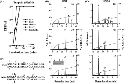
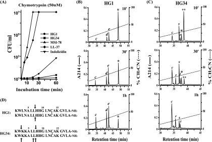
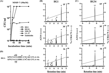
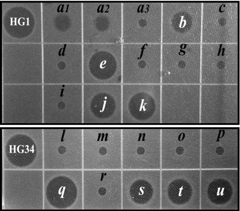
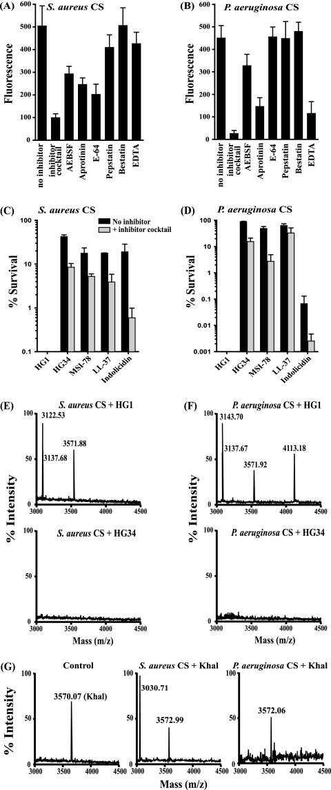
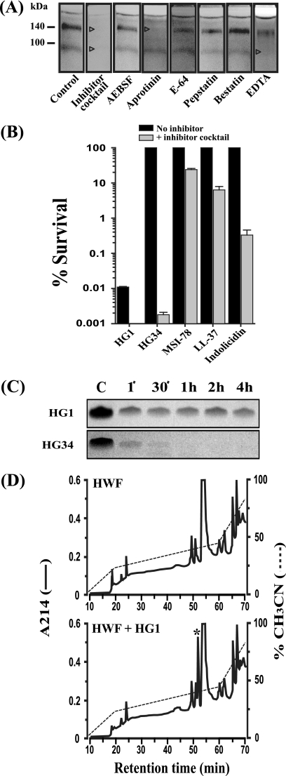
Cationic antimicrobial peptides (AMPs) have attracted a great deal of interest as a promising candidate for a novel class of antibiotics that might effectively treat recalcitrant infections caused by a variety of microbes that are resistant to currently available drugs. However, the AMPs are inherently limited in that they are inevitably susceptible to attacks by proteases generated by human and pathogenic microbes; this vulnerability severely hinders their pharmaceutical use in human therapeutic protocols. In this study, we report that a halocidin-derived AMP, designated HG1, was found to be resistant to proteolytic degradation. As a result of its unique structural features, HG1 proved capable of preserving its antimicrobial activity after incubation with trypsin, chymotrypsin, and human matrix metalloprotease 7 (MMP-7). Additionally, HG1 was observed to exhibit profound antimicrobial activity in the presence of fluid from human skin wounds or proteins extracted from the culture supernatants of Staphylococcus aureus and Pseudomonas aeruginosa. Greater understanding of the structural motifs of HG1 required for its protease resistance might provide feasible ways to solve the problems intrinsic to the development of an AMP-based antibiotic.


Figures

FIG. 1.
FIG. 1.
Effect of trypsin on the anti-MRSA activity of AMPs. (A) Each peptide was incubated with trypsin for different times and tested for its anti-MRSA activity via a colony count assay. The recovered CFU were counted and expressed in CFU/ml, and the results shown are representative of three separate experiments. (B) HPLC profiles for HG1 samples at 10, 30 min, or 1 h after incubation with trypsin. For the first 10 min after sample loading, the column was washed with solution A (H2O containing 0.01% trifluoroacetic acid [TFA]) at a flow rate of 1 ml/min. The concentration of solution B (acetonitrile containing 0.01% TFA) increased in a linear fashion to 25% over 10 min and then to 45% at 40 min. Peaks a, b, and HG1 (*) were eluted at retention times of 36.5, 43.5, and 52 min, respectively. (Inset) Analytical acid urea-PAGE for the three dimeric peptides included in peak a. Lane a, peak a; lane 1, 15-15 homodimer; lane 2, 15-14 heterodimer; lane 3, 14-14 homodimer. (C) In the case of RP-HPLC for the HG34 sample, after 10 min of washing with solution A, the acetonitrile concentration increased in a linear fashion, to 15% for 10 min and then to 35% for 40 min. Each peak was eluted at the following retention times: peak l, 22 min; peak m, 34 min; peak n, 35.5 min; peak o, 40.5 min; peak p, 41.5 min; peak q, 46 min; and HG34 (**), 49 min. (D) Arrows indicate trypsin scissile bonds on each sequence. Dashed arrows signify sites that were incompletely cleaved by the exopeptidic activity of trypsin.
FIG. 2.
FIG. 2.
Effect of chymotrypsin on anti-MRSA activity of AMPs. (A) As in the experiments for Fig. 1A, the chymotrypsin resistance of each peptide was evaluated by measuring the remaining anti-MRSA activity at different times after incubation with chymotrypsin. The results shown are also representative of three separate experiments. (B and C) At 10 min, 30 min, or 1 h after the incubation of HG1 or HG34 with chymotrypsin, each sample was subjected to RP-HPLC analysis. For the first 10 min, the column was washed with solution A. Then the solution B concentration increased in a linear fashion to 60% for 50 min. Each peak was detected at the following retention times: peak c, 34 min; peak d, 41 min; peak e, 42 min; HG1 (*), 48.5 min; peak r, 35.5 min; peak s, 36.8 min; peak t, 37.2 min; peak u, 37.5 min; and HG34 (**), 38.5 min. (D) Sites of cleavage by chymotrypsin are indicated as arrows in each sequence.
FIG. 3.
FIG. 3.
Effect of MMP-7 on anti-MRSA activity of AMPs. (A) The MMP-7 resistance of each peptide was presented by measuring the remaining anti-MRSA activity at different times after incubation with MMP-7. The results shown are also representative of three separate experiments. (B and C) At 10 min, 30 min, or 1 h after the incubation of HG1 or HG34 with MMP-7, each sample was subjected to RP-HPLC analysis under identical conditions as shown in Fig. 2B and C. Each peak was detected at the following retention times: peak f, 32.4 min; peak g, 35.9 min; peak h, 36.5 min; peak i, 37.5 min; peak j, 42.3 min; peak k, 42.6 min; HG1 (*), 49.5 min; and HG34 (**), 39 min. (D) MMP-7 cleavage sites are indicated as arrows in the amino acid sequence of HG1.
FIG. 4.
FIG. 4.
Radial diffusion assay for anti-MRSA activities of the peptide fragments generated by trypsin, chymotrypsin, or MMP-7 digestion. All peptide fragments indicated in the HPLC profiles of Fig. 1, 2, and 3 were subjected to radial diffusion assays against MRSA. Each peptide was resuspended in 0.01% acetic acid (200 μg/ml), and 5 μl (1 μg) of each sample was loaded into the well.
FIG. 5.
FIG. 5.
Resistance of AMPs against proteases secreted from S. aureus V8 or P. aeruginosa. (A and B) Proteolytic activity of the bacterial CS proteins. Each bacterial CS protein was measured with fluorescein isothiocyanate (FITC)-conjugated casein substrate. A variety of protease inhibitors were used to determine the nature of proteases: serine protease inhibitors (AEBSF and aprotinin), cysteine protease inhibitor (E-64), aspartic protease inhibitor (pepstatin), aminopeptidease inhibitor (bestatin), and metalloprotease inhibitors (EDTA). Data were obtained from three independent experiments and are expressed as mean values ± standard deviations (P < 0.05). The remaining anti-MRSA activity of AMPs was examined 1 h after incubation with proteins extracted from CS of S. aureus (C) or P. aeruginosa (D). The resistance of each peptide was evaluated in terms of % bacterial survival. The data were obtained from experiments repeated three times on different days and are expressed as mean values ± standard deviations (P < 0.05). (E, F, and G) MALDI-TOF-mass analyses conducted with HG1, HG34, or Khal samples after 1 h of incubation with the proteins of each bacterial CS.
FIG. 6.
FIG. 6.
Proteolytic stability of HG1 in the presence of human wound fluid (HWF). (A) Casein zymography analyses for the proteolytic activity of HWF. Each lane of the gel was dissected, and the sliced gels were incubated in the enzyme reaction buffer containing each of the protease inhibitors. Pictures were processed with Adobe Photoshop software. Arrowheads indicate the parts that disappeared from the gel after inhibitor treatment. (B) Each peptide was incubated with 20% HWF (solid bars) or HWF pretreated with protease inhibitor cocktail (gray bars) and then tested for the remaining anti-MRSA activity via colony count assay. As in Fig. 3A, the resistance of each peptide to HWF was evaluated by % bacterial survival. Experiments were also repeated three times, and the mean values (P < 0.05) were used to construct a graph. (C) After incubation with 20% HWF for different times, 1.5 μl was removed from each sample with a final peptide concentration of 200 μg/ml and subjected to 16.5% Tricine SDS-PAGE gel. Accordingly, 0.3 μg of HG1 or HG34 was loaded onto each lane. For a control, 1 μg of HG1 or HG34 was loaded for lane C. The gel was then stained with Coomassie blue. (D) HPLC profile for HG1 samples at 4 h after incubation with 20% HWF. HPLC was conducted under the same conditions as in the case of the trypsin-treated HG1 sample (Fig. 1B). As a result, HG1 was eluted as a distinct peak (*) at the 52-min retention time, which was not noted in the case of the control sample without HG1.

Similar articles

Cited by

References

    1. Batista, C. V., A. Scaloni, D. J. Rigden, L. R. Silva, A. Rodrigues Romero, R. Dukor, A. Sebben, F. Talamo, and C. Bloch. 2001. A novel heterodimeric antimicrobial peptide from the tree-frog Phyllomedusa distincta. FEBS Lett. 494:85-89. - PubMed
    1. Brewer, D., and G. Lajoie. 2002. Structure-based design of potent histatin analogues. Biochemistry 41:5526-5536. - PubMed
    1. Campopiano, D. J., D. J. Clarke, N. C. Polfer, P. E. Barran, R. J. Langley, J. R. Govan, A. Maxwell, and J. R. Dorin. 2004. Structure-activity relationships in defensin dimers: a novel link between beta-defensin tertiary structure and antimicrobial activity. J. Biol. Chem. 279:48671-48679. - PubMed
    1. Clinical and Laboratory Standards Institute. 2006. Methods for dilution antimicrobial susceptibility tests for bacteria that grow aerobically, 7th ed. Approved standard M7-A7. Clinical and Laboratory Standards Institute, Wayne, PA.
    1. Dalla Serra, M., O. Cirioni, R. M. Vitale, G. Renzone, M. Coraiola, A. Giacometti, C. Potrich, E. Baroni, G. Guella, M. Sanseverino, S. De Luca, G. Scalise, P. Amodeo, and A. Scaloni. 2008. Structural features of distinctin affecting peptide biological and biochemical properties. Biochemistry 47:7888-7899. - PubMed

Publication types

MeSH terms

LinkOut - more resources