. 2010 Apr 6;5(4):e10030.
doi: 10.1371/journal.pone.0010030.

Granuloma encapsulation is a key factor for containing tuberculosis infection in minipigs

Affiliations

Granuloma encapsulation is a key factor for containing tuberculosis infection in minipigs

Olga Gil et al. PLoS One. .

Abstract

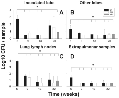
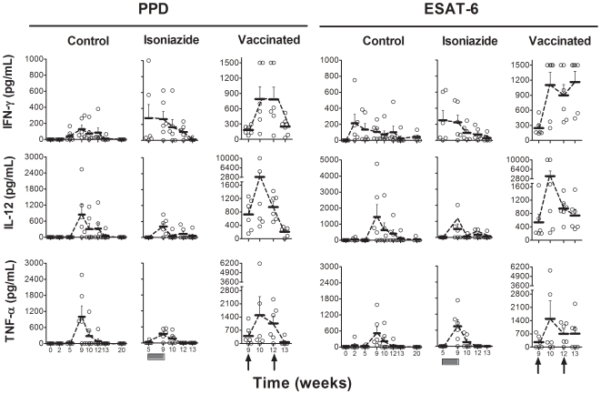
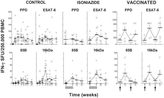
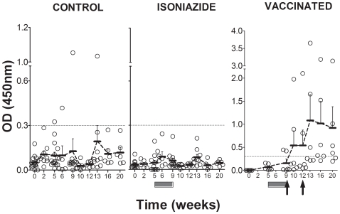
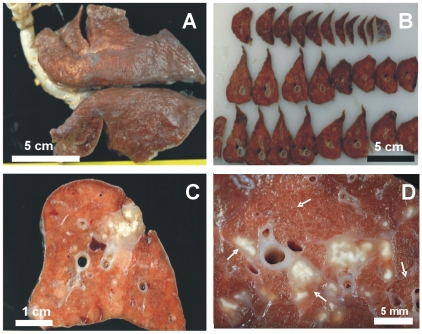
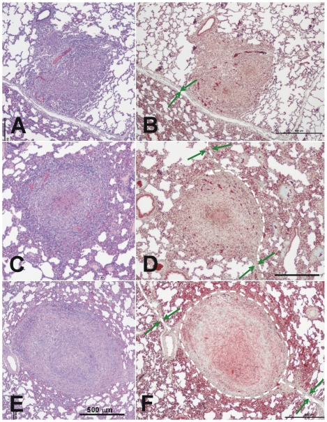
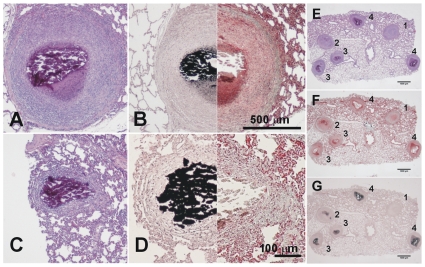
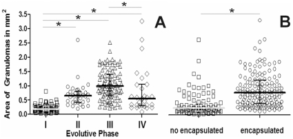
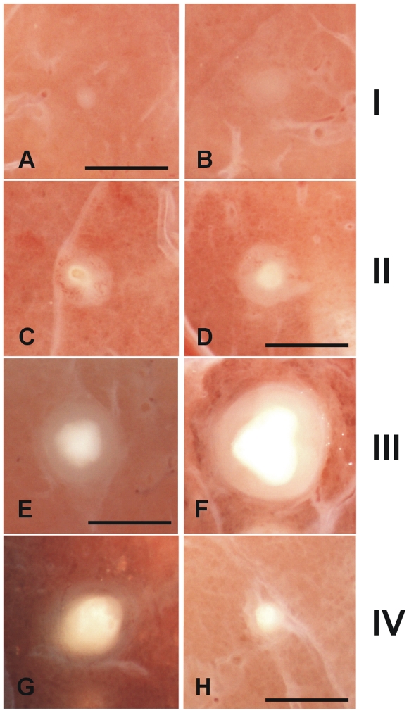
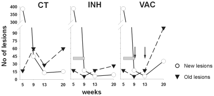
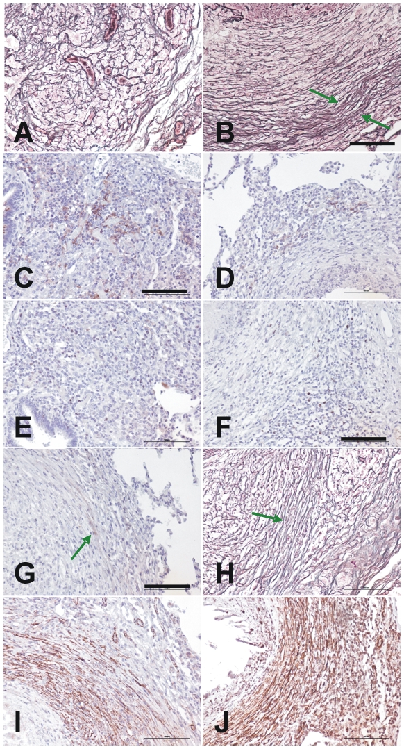
A transthoracic infection involving a low dose of Mycobacterium tuberculosis has been used to establish a new model of infection in minipigs. The 20-week monitoring period showed a marked Th1 response and poor humoral response for the whole infection. A detailed histopathological analysis was performed after slicing the formalin-fixed whole lungs of each animal. All lesions were recorded and classified according to their microscopic aspect, their relationship with the intralobular connective network and their degree of maturity in order to obtain a dissemination ratio (DR) between recent and old lesions. CFU counts and evolution of the DR with time showed that the proposed model correlated with a contained infection, decreasing from week 9 onwards. These findings suggest that the infection induces an initial Th1 response, which is followed by local fibrosis and encapsulation of the granulomas, thereby decreasing the onset of new lesions. Two therapeutic strategies were applied in order to understand how they could influence the model. Thus, chemotherapy with isoniazid alone helped to decrease the total number of lesions, despite the increase in DR after week 9, with similar kinetics to those of the control group, whereas addition of a therapeutic M. tuberculosis fragment-based vaccine after chemotherapy increased the Th1 and humoral responses, as well as the number of lesions, but decreased the DR. By providing a local pulmonary structure similar to that in humans, the mini-pig model highlights new aspects that could be key to a better understanding tuberculosis infection control in humans.


Conflict of interest statement

Competing Interests: PJC is co-inventor of the patent claiming RUTI® as a therapeutic vaccine. Regulatory approval and further clinical development is currently being undertaken by a spin-off, the biopharmaceutical company Archivel Farma S.L., in collaboration with the Institut Germans Trias i Pujol. PJC is the Scientific Director of this spin-off. The authors confirm that this does not alter their adherence to all the PLoS ONE policies on sharing data and materials.

Figures

Figure 1
Figure 1. CFU values in the lungs.
Data (mean and standard deviation) are presented by comparing an inoculated lobe with others (pictures A and B), lung lymph nodes (C), and extrapulmonary samples (D). Every sample collected weighted 2 g. approximately. * signifies a statistically significant difference in the one-way ANOVA test (p<0.005). CT  =  control group; INH  =  chemotherapy with isoniazid; VAC  =  group treated with vaccine therapy.
Figure 2
Figure 2. Evaluation of the cellular immune response by ELISA assay.
ESAT-6 and PPD-specific IFN-γ, IL-12 and TNF-α production after stimulation of PBMCs, as determined by ELISA assay. Means and standard error of the means (SEM) are drawn in gray and connected by dotted lines. The black boxes show the period of INH treatment and the black arrows the time of therapeutic vaccination.
Figure 3
Figure 3. Evaluation of the cellular immune response by ELISPOT assay.
Evaluation of ESAT-6, Ag85B, 16 kDa and PPD-specific IFN-γ production after stimulation of PBMCs, as determined by ELISPOT assay. Means and SEM are drawn in black and connected by dotted lines. The black boxes show the period of INH treatment and the black arrows the time of therapeutic vaccination.
Figure 4
Figure 4. Evaluation of the humoral response.
Humoral response against PPD is presented. The dotted line indicates the background threshold. Means and SEM are drawn in black and connected by dotted lines. The dark-grey boxes show the period of INH treatment and the black arrows the time of therapeutic vaccination.
Figure 5
Figure 5. Macroscopic pathology of the whole lung showing the intralobular septa network.
Macroscopic pathology of the whole lung (A), sliced lobes (B) and the inoculation site (C). White arrows indicate normal septa and the increased thickness of those surrounding lesions (D).
Figure 6
Figure 6. Microscopic evolution of recent lesions, showing the relationship between the granulomas and the intralobular septa.
A–D: Phase I lesions; E and F: Phase II lesions. Images A and B show the initial evolution phase where the granuloma touches an intralobular septa but there is still no fibroblast proliferation. This can be seen in images C and D, where the septa increase in thickness and start to surround the granuloma, as shown by the white dotted lines. Images E and F show how the granuloma is finally surrounded by a thick collagenic mantle. Pictures A, C and D were stained with haematoxylin and eosin (H&E), while B, D and F were stained with Masson's trichromic. The original magnification of the large images is ×40 whereas all insets, except F (x100), are magnified ×400. The green arrows show the septa and the dotted white lines the trajectory of the capsule.
Figure 7
Figure 7. Microscopic evolution of the old lesions.
Once the granuloma is structured, the necrotic process starts and calcification appears. Images A and B (phase III) show a well-structured and encapsulated granuloma with necrotic calcification. Images C and D show lesions of an advanced evolution (phase IV), with granulomas containing a large amount of calcification and fibrosis. Samples A and C were stained with H&E, whereas samples B and D were half stained with Masson's trichromic and von Kossa stain, which shows calcification in black. Pictures E to G were stained with H&E, Masson's trichromic and von Kossa stain, respectively. Lesions 1 and 2 are phase II lesions and differ only in the initial mineralization seen in lesion 2. The other lesions are all phase III lesions that have progressed differently. Original magnification is ×10.
Figure 8
Figure 8. Evolution of the area of granulomas according to their evolutive phase.
Individual data are presented in relation to the evolutive state of the lesion (A) or its encapsulation status (B). Mean and quartiles are presented in each case. Inter-group differences were determined by Dunn's One Anova on ranks test and are marked with * if statistically significant (p<0.05).
Figure 9
Figure 9. Macroscopic evolution of the lesions.
Classification of the fixed pulmonary lesions as they appear under the stereoscopic microscope. Considering the histological evolution of the lesions, and taking into account the sequential appearance of encapsulation and calcification, we have divided the lesions into four phases. Phase I is characterized by the presence of cellular infiltration (A and B). The intragranulomatous necrosis, which is characterized by the presence of an opaque zone inside the granuloma (C and D), appears during Phase II and structuration of the lesion starts. Phase III (E and F) involves the onset of calcification, which gives a shiny aspect to the central opacity, which grows in size. These lesions are characterized by the cartilaginous texture of the lesion when touched with the forceps. Phase IV lesions (G and H) are characterized by predominance of the calcification and a thin surrounding infiltration. Original magnification is ×10. Scale bar: 1 mm.
Figure 10
Figure 10. Evolution of recent and old lesions.
The results show the median of the values represented in Table 2. The gray bar shows the INH treatment period and vaccine inoculation is represented by the black arrows. CT  =  control group; INH  =  chemotherapy with isoniazid; VAC  =  group treated with vaccine therapy.
Figure 11
Figure 11. Evolution of the dissemination rate (DR).
Individual data points are represented by full or open circles in control (CT) and isoniazid-treated (INH) groups respectively, and are adjusted to an exponential regression. The continuous and dotted lines represents the adjustment for the CT (y = 5620.45−1.071·x + 0.5460) and INH (y = 295.62−0.4848·x +0.8793) groups. Both adjustments were statistically significant (p<0.0001)
Figure 12
Figure 12. Characterization of the dual fibrotic responses in granuloma evolution.
Figures A, B and H show reticulin stain of initial Phase I (A) and Phase III granulomas (the peripheral capsule is marked with a green arrow in B and H). C and D present immunostaining with anti CD10 and show an increase in the center (C) or the periphery (D) of Phase I and III granulomas, respectively. E and F also show the differences between these evolutive phases with the same proliferation pattern stained with Ki 67. G shows recognition of the capsule by anti collagen type 1 antibodies, whereas I and J show the identification of myofibroblasts using anti smooth muscle and anti HHF35 antibodies respectively. Original magnification is ×200. Scale bar: 100 µm.

References

    1. WHO. Global tuberculosis control: epidemiology, strategy, financing: WHO report 2009. 2009. Geneva, Switzerland.
    1. Parrish N, Dick J, Bishai W. Mechanisms of latency in Mycobacterium tuberculosis. Trends Microbiol. 1998;6:107–112. - PubMed
    1. Sterling TR, Bethel J, Goldberg S, Weinfurter P, Yun L, et al. The scope and impact of treatment of latent tuberculosis infection in the United States and Canada. Am J Respir Crit Care Med. 2006;173:927–931. - PubMed
    1. Bucher HC, Griffith LE, Guyatt GH, Sudre P, Naef M, et al. Isoniazid prophylaxis for tuberculosis in HIV infection: a meta-analysis of randomized controlled trials. Aids. 1999;13:501–507. - PubMed
    1. Targeted tuberculin testing and treatment of latent tuberculosis infection. This official statement of the American Thoracic Society was adopted by the ATS Board of Directors, July 1999. This is a Joint Statement of the American Thoracic Society (ATS) and the Centers for Disease Control and Prevention (CDC). This statement was endorsed by the Council of the Infectious Diseases Society of America. (IDSA), September 1999, and the sections of this statement. Am J Respir Crit Care Med. 2000;161:S221–247. - PubMed

Publication types