

Isolated non-compaction cardiomyopathy
- PMID: 20386670
- PMCID: PMC2853150
- DOI: 10.3238/arztebl.2010.0206
Isolated non-compaction cardiomyopathy
Abstract
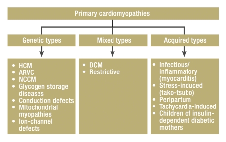
Background: Isolated non-compaction cardiomyopathy (NCCM) was first described in 1984. This disorder, a primary genetic cardiomyopathy, is now attracting increased attention.
Method: The current state of the epidemiology, pathogenesis, pathophysiology, clinical features, diagnosis, treatment, and prognosis of NCCM are discussed on the basis of a review of selected literature as well as the authors' personal experience.
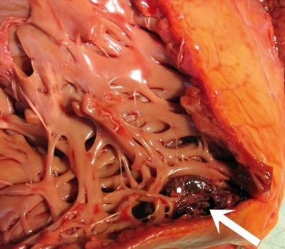
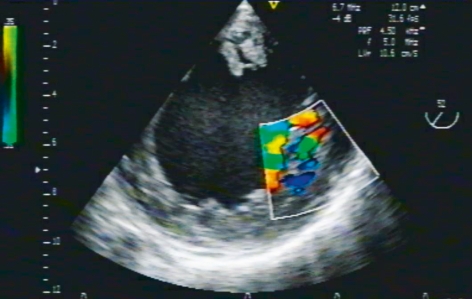
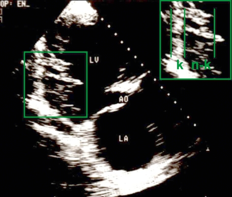
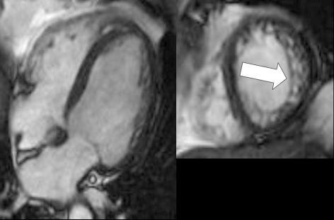
Results: The pathogenesis of NCCM is thought to involve a genetically determined disturbance of the myocardial compaction process during fetal endomyocardial morphogenesis. It is not accompanied by any other cardiac anomalies. Echocardiography is the diagnostic method of choice. The diagnosis is based on the following echocardiographic criteria: the presence of at least 4 prominent trabeculations and deep intertrabecular recesses, blood flow from the ventricular cavity into the intertrabecular recesses, and a typical bilaminar structure of the affected portion of the left ventricular myocardium. NCCM can also be diagnosed with magnetic resonance imaging of the heart. The clinical severity of NCCM is variable; its manifestations include heart failure, thromboembolic events, and arrhythmias. The treatment is symptom-based. Patients with symptomatic NCCM have a poor prognosis.
Conclusion: NCCM is a type of cardiomyopathy that was first described 25 years ago. Its molecular genetic basis is not yet fully clear, and the same is true of its diagnosis, treatment, and prognosis. Further study of these matters is needed.
Figures
References
-
- Engberding R, Bender F. Identification of a rare congenital anomaly of the myocardium by two-dimensional echocardiography: persistence of isolated myocardial sinusoids. Am J Cardiol. 1984;53:1733–1734. - PubMed
-
- Bellet S, Gouley BA. Congenital heart disease with multiple cardiac anomalies. Report of a case showing aortic atresia, fibrous scar in myocardium and embryonal sinusoidal remains. Am J med Sci. 1932;183:458–465.
-
- Dusek J, Ostádal B, Duskova M. Postnatal persistence of spongy myocardium with embryonic blood supply. Arch Pathol. 1975;99:312–317. - PubMed
-
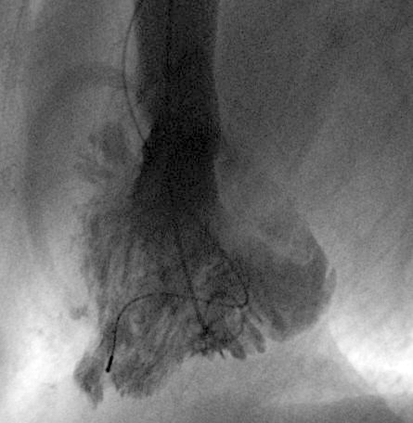
- Lauer RM, Fink HP, Petry EL, Dunn MI, Diehl AM. Angiographic demonstration of intramyocardial sinusoids in pulmonary-valve-atresia with intact ventricular septum and hypoplastic right ventricle. N Engl J Med. 1964;271:68–72. - PubMed
-
- Chin TK, Perloff JK, Williams RG, Jue K, Mohrmann R. Isolated noncompaction of left ventricular myocardium. A study of eight cases. Circulation. 1990;82:507–513. - PubMed
Publication types
MeSH terms
LinkOut - more resources
Full Text Sources