. 2010 Apr 8;6(4):e1000852.
doi: 10.1371/journal.ppat.1000852.

Exposure to HIV-1 directly impairs mucosal epithelial barrier integrity allowing microbial translocation

Affiliations

Exposure to HIV-1 directly impairs mucosal epithelial barrier integrity allowing microbial translocation

Aisha Nazli et al. PLoS Pathog. .

Abstract

While several clinical studies have shown that HIV-1 infection is associated with increased permeability of the intestinal tract, there is very little understanding of the mechanisms underlying HIV-induced impairment of mucosal barriers. Here we demonstrate that exposure to HIV-1 can directly breach the integrity of mucosal epithelial barrier, allowing translocation of virus and bacteria. Purified primary epithelial cells (EC) isolated from female genital tract and T84 intestinal cell line were grown to form polarized, confluent monolayers and exposed to HIV-1. HIV-1 X4 and R5 tropic laboratory strains and clinical isolates were seen to reduce transepithelial resistance (TER), a measure of monolayer integrity, by 30-60% following exposure for 24 hours, without affecting viability of cells. The decrease in TER correlated with disruption of tight junction proteins (claudin 1, 2, 4, occludin and ZO-1) and increased permeability. Treatment of ECs with HIV envelope protein gp120, but not HIV tat, also resulted in impairment of barrier function. Neutralization of gp120 significantly abrogated the effect of HIV. No changes to the barrier function were observed when ECs were exposed to Env defective mutant of HIV. Significant upregulation of inflammatory cytokines, including TNF-alpha, were seen in both intestinal and genital epithelial cells following exposure to HIV-1. Neutralization of TNF-alpha reversed the reduction in TERs. The disruption in barrier functions was associated with viral and bacterial translocation across the epithelial monolayers. Collectively, our data shows that mucosal epithelial cells respond directly to envelope glycoprotein of HIV-1 by upregulating inflammatory cytokines that lead to impairment of barrier functions. The increased permeability could be responsible for small but significant crossing of mucosal epithelium by virus and bacteria present in the lumen of mucosa. This mechanism could be particularly relevant to mucosal transmission of HIV-1 as well as immune activation seen in HIV-1 infected individuals.


Conflict of interest statement

The authors have declared that no competing interests exist.

Figures
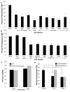
Figure 1
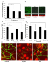
Figure 1. Primary endometrial epithelial monolayers (A) and T84 intestinal epithelial cell line (B) were exposed to 106 infectious viral units/ml of HIV-1 laboratory strains Bal, NL4-3, ADA (R5 tropic) and IIIB, MN (X4 tropic) and four clinical strains 4648 (R5 tropic) 11242, 7681 (X4 tropic) and 11249 (dual tropic).
Corresponding p24 values were as follows for R5 tropic: Bal (0.6ng/ml), ADA (two different viral stocks 0.3 ng/ml and 1040 ng/ml), 4648 (49ng/ml); X4 tropic: IIIB (0.7ng/ml and 161ng/ml), MN (103ng/ml and 1110ng/ml), NLH4-3 (93ng/ml), 7681 (773ng/ml), 11242 (342ng/ml); dual tropic 11249 (200ng/ml). Control include cultures that were mock-treated with medium without virus (mock) or virus-free supernatants (R5 and X4 control). Transepithelial resistance (TER), was measured prior to and 24 hours post- exposure to HIV-1. *p<0.001, n = 3–9. Viability of primary female genital epithelial cells and T84 intestinal epithelial cells was assessed by MTT assay 24 hours after exposure with two HIV strains IIIB (X4-tropic) and ADA (R5-tropic) and compared with mock-infected epithelial monolayers (C). The effect of apical and basolateral exposure was determined. (D). Differentiated T84 epithelial monolayers were mock infected or exposed apically or basolaterally to HIV (ADA strain, 106 infectious viral particles/ml). TER was measured at 0, 24 and 48 hours post exposure. *P<0.001, **P<0.0001, n = 3.
Figure 2
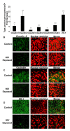
Figure 2. Confluent monolayers grown from primary endometrial epithelial cells were either mock-treated or exposed to HIV-1 (ADA strain, 106 infectious viral units/ml, p24 280ng/ml) for 8 hours.
Total RNA was extracted and cDNA was synthesized. Quantitative Real-time RT-PCR was conducted for tight junction gene expression by measuring mRNA for Claudin 1–5, Occludin and ZO-1. GAPDH, a house keeping gene, was measured for internal control (A). * p<0.01; ** p<0.001; *** p<0.0001). Immunofluorescent staining of tight junction proteins following HIV-1 exposure compared to mock-treated epithelial monolayers. Representative staining is shown for claudin-2 (B), Occludin (C), and ZO-1 (D) at 24 hours post-exposure. Magnification: 1260×. Data shown is representative of 3 separate experiments, each experiment had 3–5 replicate cultures for each experimental condition. For RNA extraction, 6–8 replicate cultures were pooled.
Figure 3
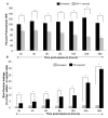
Figure 3. Primary genital epithelial monolayers were exposed to HIV-ADA (R5 strain, 106 infectious viral units/ml, p24 280ng/ml ) for 2h, 4h, 6h, 8h, 16h, 24h and 48h.
To measure barrier functions, TER was measured prior to and post exposure, ZO-1 staining was done and Dextran Blue dye leakage was measured across the monolayers at all time points. (A) TER values. p<0.001. (B) Paracellular permeability measured by addition of Blue Dextran dye on the apical side of monolayers. At different time intervals post-exposure, basolateral supernatants were sampled and absorbance was measured and compared to apical absorbance at initial time point (Time “0”). Blue Dextran leakage was calculated as a percentage of apical values. Data shown is representative of 3–4 separate experiments, each experiment had 3–5 replicate cultures for each experimental condition.
Figure 4
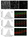
Figure 4. Genital EC monolayers were fixed after 4 or 24 hours post-HIV-1 exposure and stained for ZO-1 (green) and nucleus (red).
A–C. Series of stack planes (XY) taken through the apical extent of the monolayer. D–F. Quantification of ZO-1 (dark line) and nuclear (grey bars) staining shown as graphs. Each cell layer (1–20) corresponds to series of images from Z-stack sections taken at 1µm thickness through the cell monolayer shown on the right. X-axis illustrates cell layers from apical to basolateral. Y-axis illustrates the number of pixels present over the entire area of image. A, D. Control mock infected monolayer, 24 hours post-treatment. B,E. HIV-1 exposed monolayer, 4 hours post-treatment. C,F. HIV-1 exposed monolayer, 24 hours post-treatment. Results shown are representative of 3 separate Z-stacks collected and analyzed from each replicate, each treatment group had 3–5 replicates and the experiment was repeated 3 times. (Magnification :1260×).
Figure 5
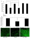
Figure 5. Primary endometrial EC monolayers were exposed to different concentrations of HIV-1 (ADA) for 24 hours.
TER values were measured, starting at viral concentration of 103 up to 107 infectious viral units/ml (equivalent to p24 values of 0.2ng–2800 ng/ml) (A).* p<0.001 **p<0.05. ZO-1 staining following exposure to different concentration of virus (B). Data shown is representative of three separate experiments, each experiment had 3–5 replicate cultures for each experimental condition.
Figure 6
Figure 6. HIV-1 induced barrier permeability is independent of viral replication.
Differentiated T84 cells were mock infected or exposed to live or UV inactivated HIV (IIIB strain, 106 infectious viral particles/ml). (A) TER values following exposure to both live and UV-inactivated HIV compared to mock treated monolayer (P<0.001). (B) ZO-1 staining in epithelial cells exposed to live and UV-inactivated HIV, but not mock infected cultures. The corresponding Z-stack series below each panel clearly shows majority of ZO-1 staining (green) in mock treated cultures on the apical side of the monolayer, while the nuclei are seen more basolaterally (red). Magnification:1260×. Primary endometrial EC monolayers (C) or intestinal T84 cells (D) were treated with either gp120 (0.1µg/ml, 0.8nM) or Tat (1.4ug/ml, 100nM) or a combination of both, for 24 hours. TER values were measured prior to and post-treatment. p<0.001, n = 6. (E) ZO-1 staining after gp120 treatment and Tat treatment. Magnification: 2520×. Data shown is representative of two (A,B) and six (C,D) separate experiments, each experiment had 3–5 replicate cultures for each experimental condition.
Figure 7
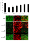
Figure 7
(A) Epithelial monolayers were treated with medium alone, HIV-1 (IIIB, 106 infectious viral particles/ml), HIV-1 in combination with gp120 neutralizing antibody (35µg/ml) or isotype control antibody (35µg/ml), gp120 or isotype antibody alone. TER measurements were taken as a measure of change in permeability and presented as percent of pre-treatment TER. p<0.01. (B) Confluent T84 epithelial cell cultures were mock infected or exposed to NL4-3 (p24, 79 ng/ml) or NL4-3 Env mutant (p24, 79 ng/ml) and TER measurements were taken prior to and 24 hours post-exposure. P<0.001 (C) ZO-1 localization after exposure to wildtype HIV-1 NL4-3 or Env NL4-3 mutant. Data shown is representative of four separate experiments, each experiment had 3–5 replicate cultures for each experimental condition.
Figure 8
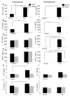
Figure 8. Primary endometrial EC and T84 intestinal monolayers were exposed to HIV-1 (ADA, 106 infectious viral units/ml, p24 280ng/ml) and apical and basolateral supernatants were collected 24 hours post-exposure and assayed by Luminex multi-analyte kit for the following cytokines: (A) TNF-α (B) IL-6, (C) IL-8, (D) MCP-1, (E) IL-10, (F) IL-1β.
*p<0.01, **p<0.001. Data shown is representative of three separate experiments from different tissues, each experiment had 3–5 replicate cultures for each experimental condition.
Figure 9
Figure 9. Primary endometrial epithelial monolayers were exposed to TNF-α or HIV-1 alone; TNF-α or HIV-1 (ADA,106 infectious viral units/ml, p24 280ng/ml) in combination with anti-TNF-α neutralizing antibody; TNF-α or HIV-1 in combination with normal mouse serum for 24 hours.
TER measurements were taken as a measure of change in permeability and presented as percentage of pre-treatment TER. Data shown is representative of two separate experiments, each experiment had 3–5 replicate cultures for each experimental condition.
Figure 10
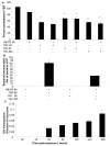
Figure 10. Bacterial and viral translocation across mucosal epithelial monolayers following HIV-1 exposure.
(A) Bacterial translocation was measured in T84 intestinal monolayers. Confluent monolayers were left untreated or treated for 6 hours with TNF-α (20ng/ml), E. coli (108 CFU/ml) , TNF-α (20ng/ml) +E. coli (108 CFU/ml), HIV-1 or HIV-1 (6 or 24 hours)+E. coli (108 CFU/ml). (A) TER measurements following various treatments in the presence or absence of E. Coli. * p<0.001. (B) Basolateral supernatants were collected and bacterial counts were done. (C) Viral translocation was determined in endometrial EC monolayers exposed to HIV-1 (ADA, 106 infectious viral units/ml, p24 280ng/ml) on the apical side. Basolateral supernatants were collected after different time intervals infectious and viral counts were done on TZM/b-l indicator cell line. Viral counts are depicted as percentage of inoculum added to the apical compartment of monolayers. Data shown is representative of two separate experiments, each experiment had 3–5 replicate cultures for each experimental condition.

Similar articles

Cited by

References

    1. Shattock RJ, Haynes BF, Pulendran B, Flores J, Esparza J. Improving defences at the portal of HIV entry: mucosal and innate immunity. PLoS Med. 2008;5:e81. doi: 10.1371/journal.pmed.0050081. - DOI - PMC - PubMed
    1. Wira CR, Grant-Tschudy KS, Crane-Godreau MA. Epithelial cells in the female reproductive tract: a central role as sentinels of immune protection. Am J Reprod Immunol. 2005;53:65–76. - PubMed
    1. Vroling AB, Fokkens WJ, van Drunen CM. How epithelial cells detect danger: aiding the immune response. Allergy. 2008;63:1110–1123. - PubMed
    1. Yu QH, Yang Q. Diversity of tight junctions (TJs) between gastrointestinal epithelial cells and their function in maintaining the mucosal barrier. Cell Biol Int. 2009;33:78–82. - PubMed
    1. Shacklett BL, Critchfield JW, Ferre AL, Hayes TL. Mucosal T-cell responses to HIV: responding at the front lines. J Intern Med. 2009;265:58–66. - PMC - PubMed

Publication types

Substances