. 2010 Apr 8;6(4):e1000898.
doi: 10.1371/journal.pgen.1000898.

Fragile x mental retardation protein regulates proliferation and differentiation of adult neural stem/progenitor cells

Affiliations

Fragile x mental retardation protein regulates proliferation and differentiation of adult neural stem/progenitor cells

Yuping Luo et al. PLoS Genet. .

Abstract

Fragile X syndrome (FXS), the most common form of inherited mental retardation, is caused by the loss of functional fragile X mental retardation protein (FMRP). FMRP is an RNA-binding protein that can regulate the translation of specific mRNAs. Adult neurogenesis, a process considered important for neuroplasticity and memory, is regulated at multiple molecular levels. In this study, we investigated whether Fmrp deficiency affects adult neurogenesis. We show that in a mouse model of fragile X syndrome, adult neurogenesis is indeed altered. The loss of Fmrp increases the proliferation and alters the fate specification of adult neural progenitor/stem cells (aNPCs). We demonstrate that Fmrp regulates the protein expression of several components critical for aNPC function, including CDK4 and GSK3beta. Dysregulation of GSK3beta led to reduced Wnt signaling pathway activity, which altered the expression of neurogenin1 and the fate specification of aNPCs. These data unveil a novel regulatory role for Fmrp and translational regulation in adult neurogenesis.


Conflict of interest statement

The authors have declared that no competing interests exist.

Figures
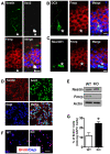
Figure 1
Figure 1. Fmrp is expressed in aNPCs and new neurons in the adult DG, and the loss of Fmrp leads to increased aNPC proliferation.
(A) Fmrp is expressed in Sox2 (white) and Nestin (green) double-positive NPCs (arrowheads) in the granule neurons of the adult hippocampus. Arrowhead points to a positive cell located at the subgranular zone adjacent to the hilar region. (B,C) Fmrp is expressed in doublecortin (DCX)-positive (B, green) and NeuroD1-postive (C, green) newly generated neurons. Asterisks identify positive cells located at the subgranular zone adjacent to the hilar region. (A–C, Fmrp, red; Dapi, blue; Scale bars = 10 µm;). (D) aNPCs cultured under proliferating conditions expressed the neural progenitor markers Nestin (cytoplasmic, red) and Sox2 (nuclear, green; Dapi in blue). (E) Proliferating WT aNPCs, but not Fmr1 KO aNPCs, expressed Fmrp. (F) Both WT and KO aNPCs incorporate the thymidine analog, BrdU, under proliferating conditions (BrdU, red; Dapi, blue; (D,F), Scale bars = 50 µm). (G) Quantitative analysis showing that a higher percentage of Fmr1 KO aNPCs incorporated BrdU. (*, p<0.05; n = 3; Student's t-test; mean ± SEM).
Figure 2
Figure 2. Loss of Fmrp leads to decreased neuronal differentiation but increased astrocyte differentiation.
(A,B) Sample immunostained cells using cell lineage markers for quantitative cell fate determination shown in (C,D). Both WT (A) and Fmr1 KO (B) aNPCs could differentiate into Tuj1+ (red) neurons and GFAP+ (green) astrocytes. (Scale bar = 50 µm; DAPI, nuclear staining, blue). (C,D) Quantitative analyses of differentiated aNPCs demonstrate that Fmr1 KO aNPCs differentiated into fewer Tuj1+ neurons (C, n = 4; p<0.01) but more GFAP+ astrocytes (D, n = 6, p<0.05). Quantification was performed using an unbiased stereology method. (E,F), Luciferase reporter assay showing that differentiating Fmr1 KO aNPCs had decreased NeuroD1 (E; n = 4, p<0.01), but increased GFAP (F, n = 3, p<0.05) promoter activities compared with WT aNPCs. A co-transfected Renilla luciferase (R-Luc) plasmid was used as a transfection control. (G,H), Real-time PCR assays showing that Fmr1 KO aNPCs had decreased NeuroD1 mRNA levels (G; n = 3, p<0.05), but increased GFAP mRNA levels (H, n = 3, p<0.05) upon differentiation. The relative mRNA levels were in comparison with GAPDH mRNA. (I,J), Acute knockdown of Fmrp expression in WT aNPCs using siRNA led to decreased neuronal differentiation (I; left NeuroD1; middle, Tuj1; right, NeuroD1-promoter), but increased astrocyte differentiation (J; left GFAP; middle, aquaporin4; right, GFAP-promoter); (K,L), Exogenously expressed WT Fmrp, but not mutant (I304N) Fmrp, could enhance neuronal differentiation (K) and repress astrocyte differentiation (L) in Fmr1 KO aNPCs. GAPDH mRNA levels were used as internal controls for real-time PCR analyses. Data are presented as mean ± SEM; *, p<0.05, **, p<0.01, ***, p<0.01, Student's t-test.
Figure 3
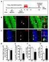
Figure 3. Loss of Fmrp alters the proliferation of neural stem and progenitor cells in vivo.
(A) Experimental scheme for assessing cell proliferation in the adult hippocampus. Cohort 1 animals had the same injection paradigm as Figure 4 and were therefore used to assess new cell survival. Cohort 2 animals were used to evaluate cell proliferation in the DG. (B) Examples of WT and Fmr1 KO brain sections stained with an antibody against BrdU (red) and DAPI (blue) for in vivo neurogenesis analyses (scale bar = 100 µm). (C) The dentate gyrus (DG) of Fmr1 KO mice exhibited increased BrdU+ cells analyzed at one day after a 7-day regimen of daily BrdU injections, suggesting increased proliferation (Cohort 1: n = 3 WT; n = 4 KO). (D) At 4 hours post-BrdU injection (6 injections within 24 hours), the number of BrdU+ cells normalized to volume of the DG was also higher in Fmr1 KO mice (p<0.05). (D–I, Cohort 2: n = 7 WT; n = 6 KO). (E) Fmr1 KO mice also had increased DG volume (size) (p<0.05). (F) Single intensity projection confocal z-series showing two different types of BrdU+ cells in the DG of the hippocampus. Upper panel, BrdU+ (red), Nestin+ (green), and GFAP− (blue) progenitor cells; Lower panel, BrdU+ (red), Nestin+ (green), and GFAP+ (blue) stem-like cells. (G) The DG of Fmr1 KO mice exhibited increased proliferation of progenitor (BrdU+ Nestin+, GFAP) cells analyzed at 4 hours following 6 BrdU injections within a 24-hour period. (H) The DG of Fmr1 KO mice exhibited increased proliferation of stem (BrdU+ Nestin+, GFAP+) cells analyzed at 4 hours after 6 BrdU injections within a 24-hour period. (n = 7 WT; n = 6 KO). Data are presented as mean ± SEM; *, p<0.05, **, p<0.01, ***, p<0.01, Student's t-test.
Figure 4
Figure 4. Loss of Fmrp alters the differentiation of neural stem and progenitor cells in vivo.
(A) Experimental scheme for assessing new cell survival and differentiation in the adult hippocampus. (B,C) Sample confocal images showing newborn cells that had differentiated into NeuN+ neurons (B, asterisk) or S100β+ astrocytes (C, asterisk). Asterisks, but not arrowheads, indicate BrdU+ cells that have differentiated into either a neuron (H) or an astrocyte (E). (D) At 4 weeks post-labeling, BrdU+ cells in the Fmr1 KO DG were no different from WT mice (n = 7 WT; n = 9 KO). (E) At 4 weeks post-BrdU injection, brains were analyzed for survival of newborn cells in the DG. The ratio of BrdU+ cells at 4 weeks post-injection (numbers used for D) over 1 day post-injection (average of BrdU+ cells shown in Figure 3D) indicated that Fmr1 KO mice had fewer surviving newborn cells in the DG. (F,G) Quantitative analysis indicated that newborn cells in Fmr1 KO mice differentiated into a lower percentage of neurons (F) but a higher percentage of astrocytes (G) compared with WT mice (n = 9 WT; n = 9 KO). All data are shown as mean ± SEM, and Student's t-test was used for all the analyses. *, p<0.05, **, p<0.01 ***, p<0.001.
Figure 5
Figure 5. Identification of the mRNAs regulated by Fmrp in aNPCs.
(A) Western blotting shows the amount of Fmrp in both input and immunoprecipitated Fmrp-containing mRNP complexes from both WT and Fmr1 KO aNPCs. (B) The RNAs from Input and from Fmrp-IP of WT and KO cells were isolated and subjected to cDNA synthesis and real-time PCR quantification. The results confirmed that Fmrp binds to the mRNAs of MAP1B, EF1a, CDK4, cyclin D1, and GSK3β in WT aNPCs. KO aNPCs and ß-Actin mRNA analyses were used as negative controls. (C) Representative western blotting image showing the protein expression levels of the target genes of Fmrp in both WT and Fmr1 KO aNPCs. EIF5 was used as a loading control for MAP1B, and ß-actin was used as a loading control for the others in western blots. Quantification of western blot band intensities is shown in Figure S6A.
Figure 6
Figure 6. Loss of Fmrp leads to a deficit in the Wnt signaling pathway and reduced Neurog1 expression in aNPCs.
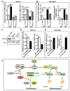
(A) In differentiating Fmr1 KO aNPCs (24 hours after initiation of differentiation), the GSK3β protein level was higher and β-catenin protein level was lower compared with differentiating WT aNPCs. (B) Differentiating Fmr1 KO aNPCs have defective Wnt signaling, as indicated by the level of TCF/LEF-luciferase activity. A mutant promoter with the TCF/LEF site mutated was used as a negative control (n = 3). (C–F) The GSK3β inhibitor SB216763 (SB) could partially rescue the reduced neuronal (C,D) and increased astrocyte (E,F) differentiation deficits of Fmr1 KO aNPCs. SB (dissolved in DMSO) was added at initiation of differentiation at 4 µM. An equal amount of DMSO was added to WT and KO control aNPCs. Cell differentiation was assessed by the relative mRNA levels of NeuroD1 (C), Tuj1 (D), GFAP (E), and aquporin4 (F). GAPDH mRNA levels were used as an internal control. (G) Real-time quantitative PCR results show that early differentiating (24 hours) WT aNPCs transiently express high levels of Neurog1 (∼10-fold induction compared with 0 hour; n = 4). This Neurog1 induction is drastically impaired in differentiating (24 hours) Fmr1 KO aNPCs (<2×-fold; n = 4). Proliferating aNPCs (0 hour), and later differentiating (48 hours) cells, expressed a minimal level of Neurog1. Inset, similar results obtained by regular RT-PCR. (H) The Wnt receptor ligand, Wnt3a, is required for activating the Neurog1 promoter during differentiation. In the presence of Wnt3a, Neurog1 promoter activity was significantly lower in Fmr1 KO aNPCs compared with WT cells. Neurog1 promoter activity was undetectable in the absence of Wnt3a (n = 3). (I) Exogenously expressed wild-type Fmr1, but not mutant Fmr1, could promote the Neurog1 transcription as assessed by Neurog1 promoter activities in both Fmr1 KO and WT aNPCs (n = 3). All data are shown as mean ± SEM, and Student's t-test was used for all the analyses. *, p<0.05, **, p<0.01***, p<0.001.
Figure 7
Figure 7. Neurog1 regulates the fate specification of aNPCs.
(A,B) Exogenously expressed Neurog1 could rescue the neuronal and astrocyte differentiation deficits of Fmr1 KO aNPCs, as assessed by real-time PCR of neuron (NeuroD1 and Tuj1) and astrocyte (GFAP and aquaporin4)-specific gene expression (n = 3; Control, pCDNA3 empty vector) (C) Neurog1-siRNA could specifically reduce the Neurog1 protein expression from a co-transfected Neurog1 expression vector. siRNA-2 was more effective at reducing Neurog1 protein expression, and was therefore used in all functional tests. NC-siRNA: Nonsilencing Control siRNA. (D,E) Acute knockdown of Neurog1 expression in aNPCs led to reduced neuronal differentiation (D), but increased astrocyte differentiation (E) in WT aNPCs, as assessed by real-time PCR of cell lineage-specific genes (n = 3). Cell differentiation was assessed by the relative mRNA levels of NeuroD1 (A and D, left), Tuj1 (A and D, right), GFAP (B and E, left), and aquporin4 (B and E, right). GAPDH mRNA levels were used as an internal control for all real-time PCR analyses, unless stated otherwise. (F) Model of Fmrp functions in adult neurogenesis. By regulating the translation of cyclin D1 and CDK4, Fmrp controls the proliferation of aNPCs. By controlling the translation of GSK3β, Fmrp maintains the proper intracellular levels of β-catenin and Wnt signaling. Upon differentiation, β-catenin positively regulates the expression of Neurog1, which promotes neuronal differentiation and represses glial differentiation. All data are shown as mean ± SEM, and Student's t-test was used for all the analyses. *, p<0.05; ***, p<0.001.

References

    1. Penagarikano O, Mulle JG, Warren ST. The pathophysiology of fragile x syndrome. Annu Rev Genomics Hum Genet. 2007;8:109–129. - PubMed
    1. Lu R, Wang H, Liang Z, Ku L, O'Donnell WT, et al. The fragile X protein controls microtubule-associated protein 1B translation and microtubule stability in brain neuron development. Proc Natl Acad Sci U S A. 2004;101:15201–15206. - PMC - PubMed
    1. Sung YJ, Dolzhanskaya N, Nolin SL, Brown T, Currie JR, et al. The fragile X mental retardation protein FMRP binds elongation factor 1A mRNA and negatively regulates its translation in vivo. J Biol Chem. 2003;278:15669–15678. - PubMed
    1. Bassell GJ, Warren ST. Fragile X syndrome: loss of local mRNA regulation alters synaptic development and function. Neuron. 2008;60:201–214. - PMC - PubMed
    1. Brennan FX, Albeck DS, Paylor R. Fmr1 knockout mice are impaired in a leverpress escape/avoidance task. Genes Brain Behav. 2006;5:467–471. - PubMed

Publication types

MeSH terms

Substances