. 2010 Apr 8;6(4):e1000904.
doi: 10.1371/journal.pgen.1000904.

Cyclic expression of lhx2 regulates hair formation

Affiliations

Cyclic expression of lhx2 regulates hair formation

Gunilla Törnqvist et al. PLoS Genet. .

Abstract

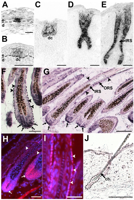
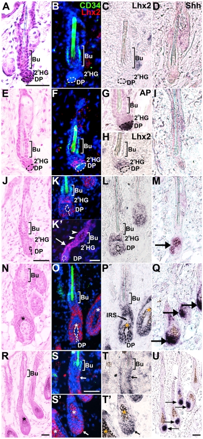
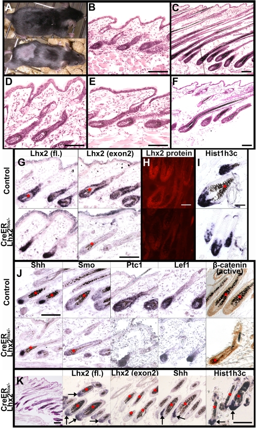
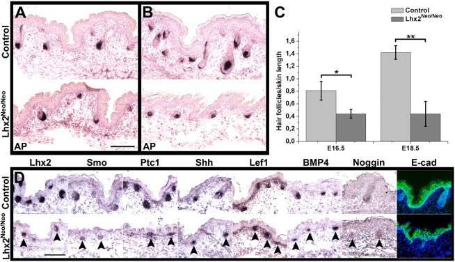
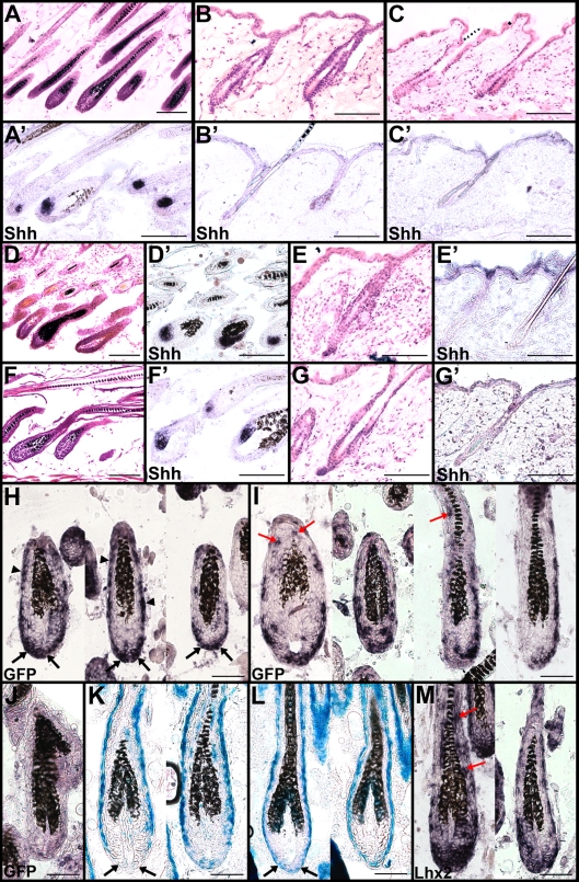
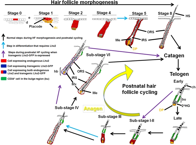
Hair is important for thermoregulation, physical protection, sensory activity, seasonal camouflage, and social interactions. Hair is generated in hair follicles (HFs) and, following morphogenesis, HFs undergo cyclic phases of active growth (anagen), regression (catagen), and inactivity (telogen) throughout life. The transcriptional regulation of this process is not well understood. We show that the transcription factor Lhx2 is expressed in cells of the outer root sheath and a subpopulation of matrix cells during both morphogenesis and anagen. As the HFs enter telogen, expression becomes undetectable and reappears prior to initiation of anagen in the secondary hair germ. In contrast to previously published results, we find that Lhx2 is primarily expressed by precursor cells outside of the bulge region where the HF stem cells are located. This developmental, stage- and cell-specific expression suggests that Lhx2 regulates the generation and regeneration of hair. In support of this hypothesis, we show that Lhx2 is required for anagen progression and HF morphogenesis. Moreover, transgenic expression of Lhx2 in postnatal HFs is sufficient to induce anagen. Thus, our results reveal an alternative interpretation of Lhx2 function in HFs compared to previously published results, since Lhx2 is periodically expressed, primarily in precursor cells distinct from those in the bulge region, and is an essential positive regulator of hair formation.


Conflict of interest statement

The authors have declared that no competing interests exist.

Figures

Figure 1
Figure 1. Lhx2 is expressed from early stages of morphogenesis and becomes restricted to the proximal part of the hair bulb and the ORS in fully developed HFs.
Lhx2 expression analysed by in situ hybridization (A–G,J) and immunohistochemistry (H-I) of tissue sections of HFs at different stages of morphogenesis (A–F) and in postnatal anagen (G–I) and telogen (J). (A) Lhx2 expression in basal keratinocytes at the pre-germ stage (Stage 0) prior to any obvious morphological change of the keratinocytes or the underlying dermis. (B) HF at the hair germ or placode stage (Stage 1) showing Lhx2 expression in the keratinocytes located at the local thickening of the epidermis. Reorganisation of mesenchymal cells in the dermis beneath the placode is indicative of formation of a dermal condensate. (C) HF at germ/peg stage (Stage 2–3) of HF morphogenesis. (D) HF at the peg stage (Stage 4), Lhx2 is expressed in the entire epithelial portion of the HF. (E) HF at the bulbous peg stage (Stage 5–6) when formation of the IRS has begun. Lhx2 is still widely expressed in outer layers of the HF and in the proximal extension the ORS in the future hair bulb whereas expression is turned off in cells in the forming IRS. (F) Fully developed HFs when the hair shaft has erupted through the epidermis (Stage 8). Lhx2 is expressed in the proximal part of the hair bulb (arrows) and in cells scattered in the ORS (arrow heads). (G) Analysis of Lhx2 expression in HFs during postnatal anagen Sub-stage VI revealing expression in the proximal part of the hair bulb (arrows) and in cells scattered in the ORS (arrow heads). (H,I) Immunohistochemical analysis of Lhx2 expression in anagen Sub-stage VI HFs revealing presence of nuclear Lhx2 protein in cells in proximal part of the hair bulb and in matrix cells (H, arrows) and in cells scattered in the ORS (H and I, arrow heads). (J) Analysis of Lhx2 expression in adult HFs in the extended telogen in 7–8 weeks old mice revealing no detectable expression. ch, club hair; dc. dermal condensate; d, dermis; e, epidermis. Scale bars, (A–I) 50 µm; (J) 100 µm.
Figure 2
Figure 2. Lhx2 expression is associated with anagen during postnatal HF cycling and is not transcribed by CD34+ bulge cells.
HFs at different Stages and Sub-stages of postnatal cycles analysed by Hematoxylin/Eosin (H/E) staining (A,E,J,N,R), CD34 immunofluorescence (B,F,K,O,S,S’), Lhx2 in situ hybridisation (B,C,F,H,K,L,O,P,S,S’,T,T’), Lhx2 immunofluorenscence (K’), Shh in situ hybridisation (D,I,M,Q,U) and alkaline phosphatase (AP) staining (G). The CD34 immunoflouresence and Lhx2 in situ hybridisation are performed on the same section and the Lhx2 in situ signal (black) has been pseudocoloured red on the DAPI stained sections (B,F,K,O,S,S’). AP staining distinguishes the DP from the secondary HG where Lhx2 is initially expressed in late telogen (F–H). Shh expression confirms the stage of the HF cycle since Shh is only expressed during anagen. (A–D) HFs in early telogen, CD34+ cells are located in the bulge (Bu) area (B) and no Lhx2 expression (B and C) or Shh expression (D) can be detected at this stage. (E–I) HFs in late telogen, CD34+ cells are located in the bulge area (F) whereas Lhx2 is expressed by cells in the secondary hair germ (2°HG) located between the bulge area and the AP+ cells in the DP (F–H). There is no overlap in Lhx2 and CD34 expression (F). Shh is not expressed confirming that the HFs are in telogen (I). (J–M) HFs in anagen Sub-stages I-II as no deposition of pigment is detected (J). CD34+ cells are located in the bulge area whereas Lhx2 is expressed in the secondary HG and the extended part of the HF enveloping the DP (K and L). Immunohistochemical analysis of Lhx2 protein reveal presence of Lhx2 in the 2°HG (K’, arrows) as well as a few cells in the lower part of the bulge region (K’, arrow heads), despite absence of Lhx2 mRNA in this part of the HF. Shh is expressed confirming that anagen has commenced (M, arrow). (N–Q) HFs in anagen Sub-stage III as deposition of pigment has started (N). CD34+ cells are located in the bulge area (O) whereas Lhx2 expression is detected in the lower transient part of the HF (O,P). Shh is expressed confirming ongoing anagen (Q, arrows). (R–U) HFs in anagen Sub-stage IV-V as the hair shaft has reached the hair canal (R). CD34+ cells are located in the bulge area whereas Lhx2 expression is detected in the proximal part of the hair bulb (S’ and T’, arrow) and in cells scattered in the ORS (S and T, arrow). Shh is expressed confirming ongoing anagen (U, arrows). * indicates melanin deposition. Scale bars, 50 µm. 2°HG, secondary hair germ; Bu, bulge; DP, dermal papilla; IRS, inner root sheath.
Figure 3
Figure 3. Lhx2 is required for anagen progression.
(A) A Lhx2flox/- control mouse (upper panel) and a CreER:Lhx2flox/- mouse (lower panel) that were shaved and treated with Tx during the first postnatal telogen and analysed during late first postnatal anagen. Hair have re-grown on the shaved area in the control mouse but not in the CreER:Lhx2flox/- mouse. (B,C) H/E staining of sections of skin from Tx-treated control mice in early anagen (B, Sub-stage III) and late anagen (C, Sub-stage VI). (D–F) H/E staining of sections of skin from Tx-treated CreER:Lhx2flox/- mice within the Tx-treated area (D,E) and outside of the Tx-treated area (F). Mutant HFs initiated anagen and developed to Sub-stage III similar to control mice (D, early anagen), but these HFs were arrested at this stage and were unable to assemble a normal hair shaft (E, late anagen). Hair shafts developed in HFs outside of the Tx-treated area in the CreER:Lhx2flox/- mice (F). (G) In situ hybridization analyses of Lhx2 expression in HFs in anagen in Tx-treated control animals (upper panels) and CreER:Lhx2flox/- animals (lower panels) using a full length (fl) Lhx2 cDNA probe (left panels) or a probe restricted to exon 2 (right panels). Control HFs revealed hybridization to both probes whereas the mutated HFs only hybridized to the full length probe confirming the expression of the truncated Lhx2 mRNA in the HFs where Lhx2 has been conditionally inactivated. (H) Immunohistochemical analysis of Lhx2 expression using and anti-Lhx2 antibody in control HFs (upper panel) and in HFs where Lhx2 has been conditionally inactivated (lower panel). (I) In situ hybridization analysis of expression of the S-phase-specific gene His1h3c in control anagen HFs (upper panel) and in anagen HFs where Lhx2 has been conditionally inactivated (lower panel). (J) Comparison of gene expression using in situ hybridization of the indicated genes between control anagen HFs (upper panel) and anagen HFs where Lhx2 has been conditionally inactivated (lower panel). Comparison of accumulation of the activated form of β-catenin (brown staining) in control HFs (upper right panel) and in HFs where Lhx2 has been inactivated (lower right panel) using an antibody specific for the dephosphorylated form of β-catenin. (K) Morphological and gene expression analyses of a Tx-treated CreER:Lhx2flox/- mouse that have not re-grown the hair at 9 week of age. H/E staining of sections of skin revealing that normal hair shafts are not assembled (left panel, compare to control skin in C). Cells in the mutant HFs express Lhx2 mRNA that hybridize to the full length Lhx2 probe (fl. arrows) but not the exon-2 specific probe revealing that the Lhx2 gene is completely inactivated. Cells in mutant HFs also express the anagen-specific gene Shh (arrows) and the S-phase-specific gene His1h3c revealing the presence of proliferating cells (arrows). * indicates melanin deposition. Scale bars 100 µm.
Figure 4
Figure 4. Reduced expression of Lhx2 hampers HF morphogenesis.
(A,B) AP staining of skin sections of E16.5 embryos (A) and E18.5 embryos (B) revealing the number of HFs in skin of control embryos (upper panels) compared to skin in Lhx2Neo/Neo embryos (lower panels). (C) Estimation of HF density based on the AP staining of skin of E16.5 embryos (left) and E18.5 embryos (right) comparing control embryos to Lhx2Neo/Neo embryos. Data are presented as average ± SD. *p<0.005. **p<0.0001 (Student’s t-test). (D) Skin sections of E16.5 embryos were analysed by in situ hybridization for expression of the indicated genes encoding mediators of signaling pathways involved in HF morphogenesis (arrow heads), and analysed by immunohistochemistry for expression of E-cad in epidermis comparing control embryos (upper panels) to Lhx2Neo/Neo embryos (lower panels). Scale bars, 100 µm.
Figure 5
Figure 5. Lhx2 expression is sufficient to induce the anagen stage of the HF cycle.
(A,A’) H/E staining of back skin HFs in anagen in 9 weeks old Tx-treated Z/Lhx2-GFP:CreER mice (A) revealing Shh expression (A’). (B,B’) H/E staining of back skin HFs in telogen in 9 weeks old Tx-treated control mice (B) revealing no Shh expression (B’). (C,C’) H/E staining of back skin HFs in telogen (C) retrieved from an area outside of the Tx-treated area in Z/Lhx2-GFP:CreER mice revealing no Shh expression (C’). (D–G,D’–G’) Representative examples of back skin HFs in 8 week and 2 day old Tx-treated Z/Lhx2-GFP:CreER mice where HFs in one individual are in anagen (D,D’) and in telogen in another individual (E,E’) showing that these mice also could enter telogen at the same time as the control animals (G,G’). The HFs were in anagen also in a slightly older Tx-treated Z/Lhx2-GFP:CreER animal (8-week- and 4-day-old) (F,F’). (H–J) In situ hybridization analyses for GFP expression in HFs from Tx-treated Z/Lhx2-GFP:CreER animals (H,I) and Z/Lhx2-GFP control mice (J). Most common expression pattern of GFP is in the proximal part of the hair bulb (H, black arrows) and in the ORS (H, arrow heads). GFP expression was also observed frequently in cells in the IRS (I, red arrows). (K,L) β-Gal staining of HFs in a Tx-treated Z/Lhx2-GFP:CreER (K) compared to HFs in a Z/Lhx2-GFP control animal (L). Lack of β-Gal+ cells in the proximal part of the hair bulb is in agreement with that most of the GFP+ cells are located at this part of the HF (black arrows). Control HFs contain numerous β-Gal+ cells in this area (L, black arrows). (M) Expression of Lhx2 analysed by in situ hybridization in HFs in Tx-treated Z/Lhx2-GFP:CreER mice. Expression is detected in all HFs and also in the IRS where Lhx2 is not expressed in control animals (red arrows). Scale bars: (A–G,A’–G’) 100 µm, (H–M) 50 µm.
Figure 6
Figure 6. Overview of the expression pattern of Lhx2 and a model for the function of Lhx2 in HF morphogenesis and postnatal HF cycling.
Lhx2 is expressed from an early time point during HF morphogenesis in basal keratinocytes before a dermal condensate (dc) is formed (Stage 0), and is broadly expressed in the epithelial part of the HF in the subsequent stages of morphogenesis. In a fully developed HF (Stage 8 of morphogenesis), Lhx2 is expressed by cells in the ORS and in the most proximal part of the hair bulb (hb). When the HF enters telogen Lhx2 expression becomes undetectable (Telogen, Early) but expression reappears in CD34 cells in the secondary hair germ (2°HG) immediately prior to anagen induction (Telogen, Late). The pattern of Lhx2 expression during morphogenesis is thereafter reiterated during anagen progression (Sub-stages I-VI) until the next anagen-catagen-telogen transition occurs and Lhx2 expression is turned off. The conditional inactivation of Lhx2 in postnatal HFs reveal that these HFs do not develop beyond Sub-stage III of the anagen phase showing that Lhx2 is required to develop further (blue arrow in anagen progression). The mouse strain homozygous for the hypomorphic allele of Lhx2 (Lhx2Neo/Neo), revealed that embryonic HFs are developmentally arrested at an early stage in this loss-of-function model. Since we rarely identified HFs at Stage 5 we suggest that Lhx2 is required to develop beyond Stage 4 (blue arrow in HF morphogenesis). HFs expressing transgenic Lhx2 (Lhx2-GFP) during postnatal HF cycling can enter catagen and telogen similar to control HFs, but transgenic Lhx2 expression causes premature initiation of anagen (purple arrows). The cells expressing transgenic Lhx2 preferentially locate to the proximal part of the hair bulb where the cells expressing endogenous Lhx2 are also located. Since transgenic expression of Lhx2 appears to induce endogenous Lhx2 expression, we suggest that numerous cells co-express the transgene and the endogenous gene, particularly in the proximal part of hb. Cells expressing transgenic Lhx2 are also located in the IRS whereas endogenous Lhx2 is not expressed in this area. 2°HG, secondary hair germ; bu, bulge region; ch, club hair; d, dermis; dc, dermal condensate; DP, dermal papilla; e, epidermis; hb, hair bulb; HS, hair shaft; IRS, inner root sheath; Me, melanocytes synthesising pigment; Mx, matrix; ORS, outer root sheath.

References

    1. Davidson P, Hardy M. The development of mouse vibrissae in vivo and in vitro. J Anat. 1952;86:342–356. - PMC - PubMed
    1. Hardy M. The secret life of the hair follicle. Trends Genet. 1992;8:55–61. - PubMed
    1. Botchkarev V, Paus R. Molecular biology of hair morphogenesis: development and cycling. J Exp Zoolog B Mol Dev Evol. 2003;298:164–180. - PubMed
    1. Fuchs E, Merrill B, Jamora C, DasGupta R. At the roots of a never-ending cycle. Develop Cell. 2001;1:13–25. - PubMed
    1. Cotsarelis G, Sun T, Lavker R. Label-retaining cells reside in the bulge area of pilosebaceous unit: implications for follicular stem cells, hair cycle, and skin carcinogenesis. Cell. 1990;61:1329–1337. - PubMed

Publication types