. 2010 Sep-Oct;16(9-10):343-51.
doi: 10.2119/molmed.2010.00031. Epub 2010 Apr 9.

Immunomodulatory drugs regulate HMGB1 release from activated human monocytes

Affiliations

Immunomodulatory drugs regulate HMGB1 release from activated human monocytes

Hanna Schierbeck et al. Mol Med. 2010 Sep-Oct.

Abstract

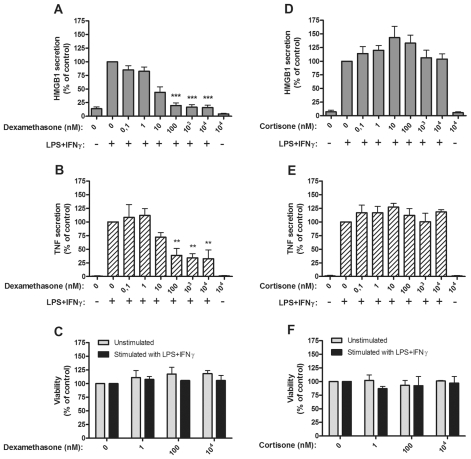
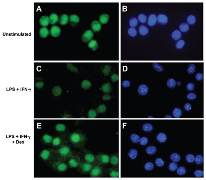
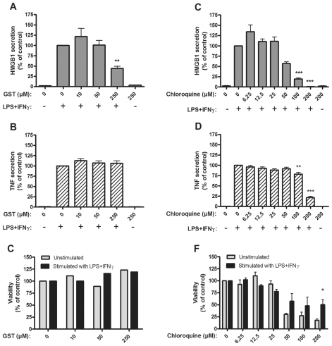
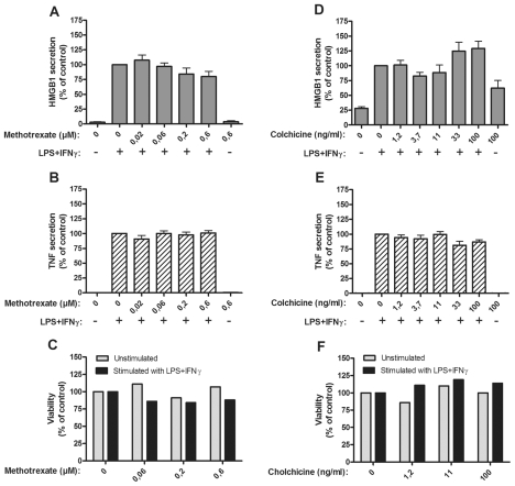
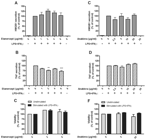
Several HMGB1-specific antagonists have provided beneficial results in multiple models of inflammatory disease-preclinical trials including arthritis. Since no HMGB1-specific targeted therapy has yet reached the clinic, we have performed in vitro studies to investigate whether any of a selection of well-established antirheumatic drugs inhibit HMGB1 release as part of its mode of action. Freshly purified peripheral blood monocytes from healthy donors were stimulated in cultures with LPS and IFNγ to cause HMGB1 and TNF release detected in ELISPOT assays. Effects on the secretion were assessed in cultures supplemented with dexamethasone, cortisone, chloroquine, gold sodium thiomalate, methotrexate, colchicine, etanercept or anakinra. Pharmacologically relevant doses of dexamethasone, gold sodium thiomalate and chloroquine inhibited the extracellular release of HMGB1 in a dose-dependent mode. Immunostaining demonstrated that dexamethasone caused intracellular HMGB1 retention. No effects on HMGB1 secretion were observed in cultures with activated monocytes by any of the other studied agents. TNF production in LPS/IFNγ-activated monocytes was readily downregulated by dexamethasone and, to some extent, by chloroquine and etanercept. We conclude that dexamethasone, gold sodium thiomalate and chloroquine share a capacity to inhibit HMGB1 release from activated monocytes.


Figures

Figure 1
Figure 1
Dexamethasone, but not cortisone, inhibited HMGB1 and TNF release from human monocytes. Effects of dexamethasone on HMGB1 and TNF release and on cell viability (A–C), and effects of cortisone on HMGB1 or TNF release and cell viability (D–F), are demonstrated. Human primary monocytes were pretreated with dexamethasone (A, B) or cortisone (D, E) for 1 h and subsequently stimulated with LPS/IFNγ for 24 or 7 h. Secretion of HMGB1 and TNF was detected by ELISPOT. Data from at least three experiments were normalized by denoting the number of spots from LPS/IFNγ stimulated cells as 100% and subsequently calculating the effect achieved by addition of the drug; P values were calculated by Kruskal–Wallis nonparametric ANOVA test. *P < 0.05; **P < 0.01; ***P < 0.001. Cell viability was assessed by Annexin-V staining (C, F).
Figure 2
Figure 2
Dexamethasone generated intracellular HMGB1 accumulation in RAW 264.7 cells activated by LPS/IFNγ. RAW 264.7 cells monocytic cell line cells were cultured for 24 h without exogenous stimulus (A, B), or with LPS/IFNγ (C, D) or with LPS/IFNγ plus 103 nmol/L dexamethasone (E, F). The cells were then fixed and stained by immunofluorescence to identify HMGB1 (green) or cell nuclei (hoechst blue). HMGB1 was dominantly expressed intranuclearly in unstimulated cells with some cells expressing additional cytoplasmic HMGB1 (A). Cells activated by LPS/IFNγ demonstrated much weaker HMGB1 signals both in the nucleus and cytoplasm (C), compared with cells in panels A and E. Cells activated by LPS/IFNγ in the presence of dexamethasone expressed the strongest cytoplasmic staining (E) of the three studied cell cultures and the intensity of the nuclear signal was at least equal to that in unstimulated cells and considerably stronger than in activated cells cultured without dexamethasone (C).
Figure 3
Figure 3
Gold sodium thiomalate and chloroquine inhibited HMGB1 release, but only chloroquine inhibited TNF release. Effects of gold sodium thiomalate (GST) on HMGB1 and TNF release and on cell viability (A–C), and effects of chloroquine on HMGB1 or TNF release and on cell viability (D–F). Human primary monocytes were pretreated with GST or chloroquine for 1 h and subsequently stimulated with LPS/IFNγ for 24 or 7 h. Secretion of HMGB1 and TNF was detected by ELISPOT. Data from at least three experiments were normalized by denoting the number of spots from LPS/IFNγ stimulated cells as 100% and subsequently calculating the effect achieved by addition of the drug; P values were calculated by Kruskal–Wallis nonparametric ANOVA test. **P < 0.01; ***P < 0.001. Cell viability was assessed by Annexin-V staining.
Figure 4
Figure 4
Methotrexate or colchicine had no effect on HMGB1 and TNF release. Effects of methotrexate on HMGB1 and TNF release and on cell viability (A–C), and effects of colchicine on HMGB1 and TNF release and on cell viability (D–F), are demonstrated. Human primary monocytes were pretreated with methotrexate or colchicine for 1 h and subsequently stimulated with LPS/IFNγ for 24 or 7 h. Secretion of HMGB1 and TNF was detected by ELISPOT. Data from at least three experiments were normalized by denoting the number of spots from LPS/IFNγ stimulated cells as 100% and subsequently calculating the effect achieved by addition of the drug; P values were calculated by Kruskal–Wallis non-parametric ANOVA test and found to be nonsignificant. Cell viability was assessed by Annexin-V staining.
Figure 5
Figure 5
Blockade of TNF or IL-1β did not suppress HMGB1 release. Effects of soluble TNF receptor (etanercept) on HMGB1 and TNF release and on cell viability (A–C), and effects of IL-1RA (anakinra) on HMGB1 and TNF release and on cell viability (D–F), are demonstrated. Human primary monocytes were pretreated with etanercept or anakinra for 1 h and stimulated with LPS/IFNγ for 24 or 7 h. Secretion of HMGB1 and TNF was detected by ELISPOT. Data from at least three experiments were normalized by denoting the number of spots from LPS/IFNγ stimulated cells as 100% and subsequently calculating the effect achieved by addition of the drug; P values were calculated by Kruskal–Wallis nonparametric ANOVA test and found to be nonsignificant. **P < 0.01; ***P < 0.001. Cell viability was assessed by Annexin-V staining.

References

    1. Yang H, Tracey KJ. Targeting HMGB1 in inflammation. Biochim. Biophys. Acta. 1799:149–56. - PMC - PubMed
    1. Andersson U, Harris HE. The role of HMGB1 in the pathogenesis of rheumatic disease. Biochim. Biophys. Acta. 1799:141–8. - PubMed
    1. Semino C, Angelini G, Poggi A, Rubartelli A. NK/iDC interaction results in IL-18 secretion by DCs at the synaptic cleft followed by NK cell activation and release of the DC maturation factor HMGB1. Blood. 2005;106:609–16. - PubMed
    1. Wang H, et al. HMG-1 as a late mediator of endotoxin lethality in mice. Science. 1999;285:248–51. - PubMed
    1. Wang H, et al. Proinflammatory cytokines (tumor necrosis factor and interleukin 1) stimulate release of high mobility group protein-1 by pituicytes. Surgery. 1999;126:389–92. - PubMed

Publication types

MeSH terms

LinkOut - more resources