. 2010 Aug;15(6):879-88.
doi: 10.1007/s00775-010-0651-0. Epub 2010 Apr 13.

Structural features promoting dioxygen production by Dechloromonas aromatica chlorite dismutase

Affiliations

Structural features promoting dioxygen production by Dechloromonas aromatica chlorite dismutase

Brandon R Goblirsch et al. J Biol Inorg Chem. 2010 Aug.

Abstract

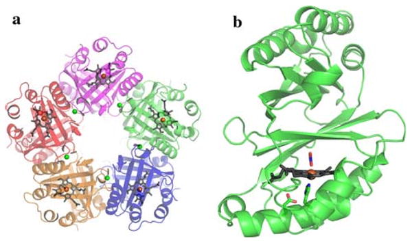
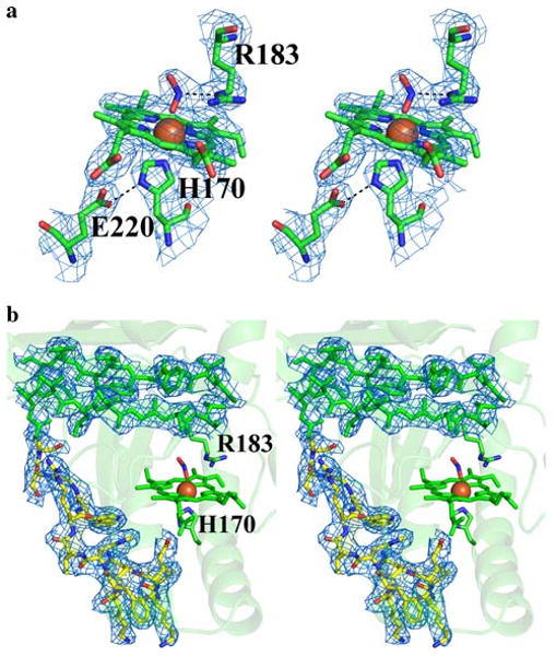
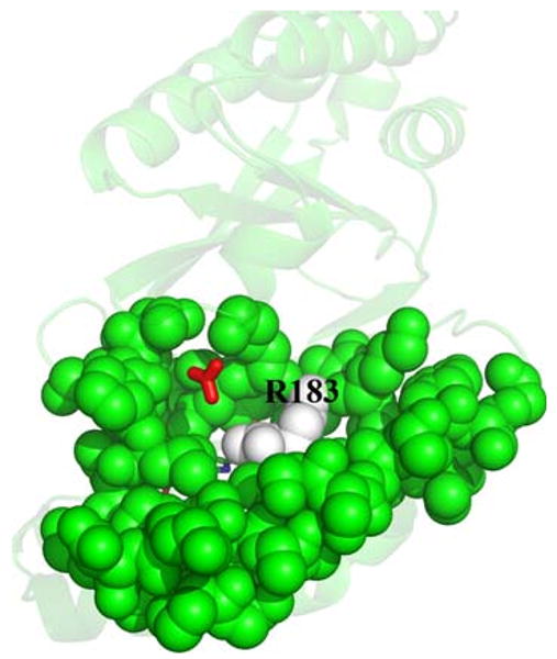
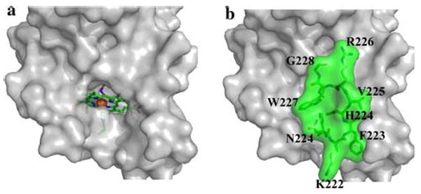
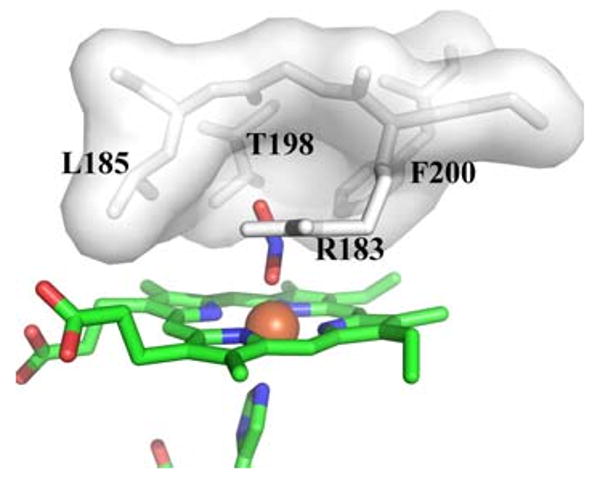
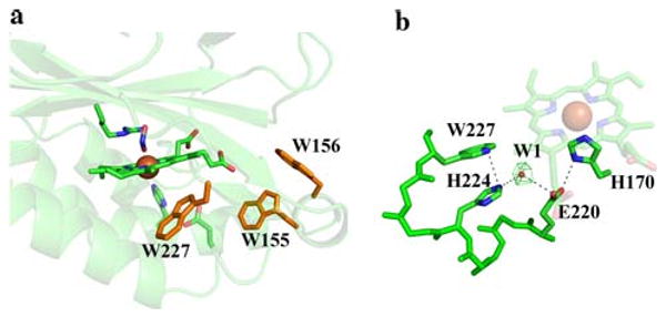
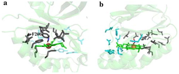
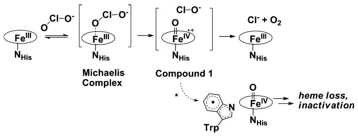
Chlorite dismutase (Cld) is a heme enzyme capable of rapidly and selectively decomposing chlorite (ClO(2) (-)) to Cl(-) and O(2). The ability of Cld to promote O(2) formation from ClO(2) (-) is unusual. Heme enzymes generally utilize ClO(2) (-) as an oxidant for reactions such as oxygen atom transfer to, or halogenation of, a second substrate. The X-ray crystal structure of Dechloromonas aromatica Cld co-crystallized with the substrate analogue nitrite (NO(2) (-)) was determined to investigate features responsible for this novel reactivity. The enzyme active site contains a single b-type heme coordinated by a proximal histidine residue. Structural analysis identified a glutamate residue hydrogen-bonded to the heme proximal histidine that may stabilize reactive heme species. A solvent-exposed arginine residue likely gates substrate entry to a tightly confined distal pocket. On the basis of the proposed mechanism of Cld, initial reaction of ClO(2) (-) within the distal pocket generates hypochlorite (ClO(-)) and a compound I intermediate. The sterically restrictive distal pocket probably facilitates the rapid rebound of ClO(-) with compound I forming the Cl(-) and O(2) products. Common to other heme enzymes, Cld is inactivated after a finite number of turnovers, potentially via the observed formation of an off-pathway tryptophanyl radical species through electron migration to compound I. Three tryptophan residues of Cld have been identified as candidates for this off-pathway radical. Finally, a juxtaposition of hydrophobic residues between the distal pocket and the enzyme surface suggests O(2) may have a preferential direction for exiting the active site.


Figures

Fig. 1
Fig. 1
Overview of the chlorite dismutase (Cld) crystal structure. a The pentamer structure. b Cld monomer. Monomers shown as a cartoon with heme drawn in gray sticks, and the iron center represented as an orange sphere. Calcium ions are shown as green spheres. Nitrite and relevant residues are colored by atom (carbon, green). This figure was generated using PyMOL (http://www.pymol.org/)
Fig. 2
Fig. 2
Stereo images of Cld 2FoFc electron density. The blue mesh represents the 20-fold-averaged 2FoFc map contoured at 3.0σ. a Cld active site at pH 6.5. Nitrite is modeled as a direct ligand bound end-on to the heme (nitrito ligation). The model is drawn as sticks and colored by atom (carbon, green). b The newly modeled residues 218–230 drawn as sticks and colored by atom (carbon, yellow). In addition, part of a β-sheet is displayed to show the general quality of the fit between the electron density and the model. The model is drawn as sticks and colored green. Iron is drawn as an orange sphere. The heme porphyrin is drawn as sticks and colored green. Arg-183 and His-170 are drawn as sticks and colored by atom (carbon, green). This figure was generated using PyMOL (http://www.pymol.org/)
Fig. 3
Fig. 3
Proposed Arg-183 gate. Surface residues close to the heme active site are drawn as solvent-accessible green spheres except for Arg-183, which is drawn as white spheres. The heme cofactor and bound nitrite are drawn as sticks colored by atom, and lie directly behind Arg-183. A hydrogen carbonate molecule was superimposed from the Azospira oryzae Cld model (Protein Data Bank entry 2vxh) [23] onto the Dechloromonas aromatica Cld model using COOT [40] and is drawn as red sticks. This figure was generated using PyMOL (http://www.pymol.org/)
Fig. 4
Fig. 4
Solvent-sequestered Cld active site. a Solvent-accessible surface of Cld without residues 221–228. b Solvent-accessible surface of the D. aromatica Cld crystal structure, including residues 221–228 (green) that are defined in D. aromatica Cld. The orientation for a and b is identical. Residues surrounding the active site are shown as a gray surface, except residues 221–228, explicitly drawn as green sticks with a green surface. The heme proximal ligand, His-170, and distally bound nitrito ligand are shown as sticks, and the iron center of the heme is shown as an orange sphere. This figure was generated using PyMOL (http://www.pymol.org/)
Fig. 5
Fig. 5
Distal pocket of Cld. Residues lining the distal pocket are shown as white sticks with a transparent surface, except for Arg-183. The heme and ligands are drawn as sticks colored by atom (carbon, green), with the iron as an orange sphere. This figure was generated using PyMOL (http://www.pymol.org/)
Fig. 6
Fig. 6
Possible sites for a tryptophan radical. a Three tryptophan residues that might be involved in radical formation are shown as orange sticks. The heme, ligands, and Arg-183 are drawn as sticks colored by atom (carbon, green), with the iron as an orange sphere. b Possible electron relay involving Trp-227. The green mesh represents a 20-fold-averaged FoFc peak at 11σ modeled as water shown as a red sphere. The heme and residues are drawn as sticks colored by atom (carbon, green), with the iron as an orange sphere. This figure was generated using PyMOL (http://www.pymol.org/)
Fig. 7
Fig. 7
Putative O2 exit direction. a Polar residues are drawn in cyan sticks and nonpolar residues are drawn as gray sticks. The heme is colored green, the nitrito ligand is in atom colors, and the iron is shown as an orange sphere. b A 90° rotation about an axis perpendicular to the heme plane. Coloring as in a. This figure was generated using PyMOL (http://www.pymol.org/)
Scheme 1
Scheme 1
Proposed mechanism of action of chlorite dismutase. The asterisk denotes the off-pathway formation of the tryptophanyl radical

References

    1. Liu S, Suflita JM. Trends Biotechnol. 1993;11:344–352. - PubMed
    1. Wackett LP. J Biol Chem. 2004;279:41259–41262. - PubMed
    1. Wackett LP, Hershberger CD. Biocatalysis and biodegradation : microbial transformation of organic compounds. ASM Press; Washington: 2001.
    1. Oremland RS, Kulp TR, Blum JS, Hoeft SE, Baesman S, Miller LG, Stolz JF. Science. 2005;308:1305–1308. - PubMed
    1. Oremland RS, Stolz JF. Science. 2003;300:939–944. - PubMed

Publication types