. 2010 Sep;11(3):407-18.
doi: 10.1007/s10162-010-0211-x. Epub 2010 Apr 13.

CD44 is a marker for the outer pillar cells in the early postnatal mouse inner ear

Affiliations

CD44 is a marker for the outer pillar cells in the early postnatal mouse inner ear

Ronna Hertzano et al. J Assoc Res Otolaryngol. 2010 Sep.

Abstract

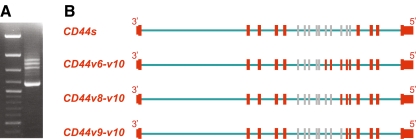
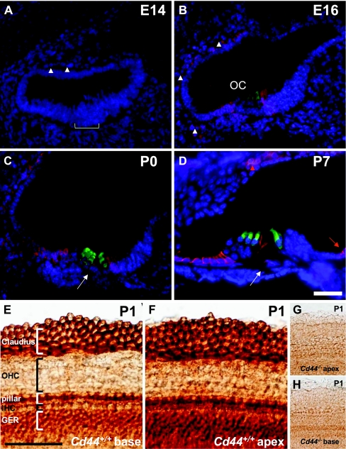
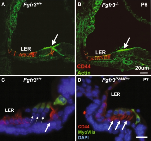
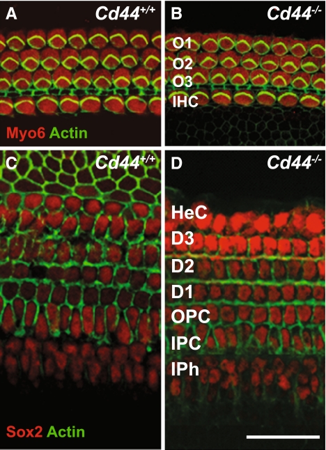
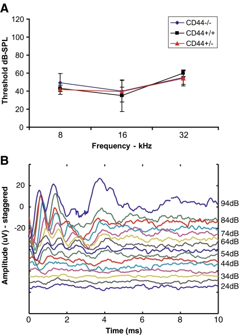
Cluster of differentiation antigens (CD proteins) are classically used as immune cell markers. However, their expression within the inner ear is still largely undefined. In this study, we explored the possibility that specific CD proteins might be useful for defining inner ear cell populations. mRNA expression profiling of microdissected auditory and vestibular sensory epithelia revealed 107 CD genes as expressed in the early postnatal mouse inner ear. The expression of 68 CD genes was validated with real-time RT-PCR using RNA extracted from microdissected sensory epithelia of cochleae, utricles, saccules, and cristae of newborn mice. Specifically, CD44 was identified as preferentially expressed in the auditory sensory epithelium. Immunohistochemistry revealed that within the early postnatal organ of Corti, the expression of CD44 is restricted to outer pillar cells. In order to confirm and expand this finding, we characterized the expression of CD44 in two different strains of mice with loss- and gain-of-function mutations in Fgfr3 which encodes a receptor for FGF8 that is essential for pillar cell development. We found that the expression of CD44 is abolished from the immature pillar cells in homozygous Fgfr3 knockout mice. In contrast, both the outer pillar cells and the aberrant Deiters' cells in the Fgfr3 ( P244R/ ) (+) mice express CD44. The deafness phenotype segregating in DFNB51 families maps to a linkage interval that includes CD44. To study the potential role of CD44 in hearing, we characterized the auditory system of CD44 knockout mice and sequenced the entire open reading frame of CD44 of affected members of DFNB51 families. Our results suggest that CD44 does not underlie the deafness phenotype of the DFNB51 families. Finally, our study reveals multiple potential new cell type-specific markers in the mouse inner ear and identifies a new marker for outer pillar cells.


Figures

FIG. 1
FIG. 1
Relative quantification of 68 CD genes in the newborn mouse inner ear using real-time RT-PCR. All results were quantified relative to the mean expression level in the samples from the cochlear sensory epithelium. The expression of CD44 and CD333 (marked with red circles) was greater than tenfold higher in the auditory sensory epithelium compared with the individual vestibular sensory epithelia. Error bars represent one standard deviation. The numerical values for the relative expression results are available in ESM Table S2.
FIG. 2
FIG. 2
CD44 isoforms in the newborn mouse cochlea. PCR amplification with CD44-specific primers identified four predominant isoforms expressed in the mouse cochlea (A). Left lane DNA ladder. Isoform sizes: 1,155, 1,447, 1,549 and 1,834 bp. Cloning and direct sequencing of the PCR products identified isoforms CD44s, CD44v6-v10, CD44v8-v10, and CD44v9-v10 as the predominant CD44 transcripts expressed in the auditory sensory epithelium (B).
FIG. 3
FIG. 3
CD44 expression in the developing mouse inner ear. Cross-sections of E14 (A), E16 (B), P0 (C), and P7 (D) wild-type mouse inner ears immunolabeled with CD44 (red) and myosin VI (green) and counterstained with DAPI (blue). CD44 is weakly expressed in some of the mesenchyme surrounding the cochlear duct at E14 (arrowheads; the prospective organ of Corti is marked with a bracket). By E16, the expression of CD44 is increased in the mesenchymal tissue (arrowheads) and is present also in the epithelium of the cochlear duct. Of note, at E16, CD44 expression is not detected in the hair cells, or in the Deiters’, Hensen’s, or pillar cells. By P0, the expression of CD44 includes the outer pillar cell (arrow). At P7, the expression of CD44 is decreased in the underlying mesenchyme and increases in intensity in Claudius and the outer pillar cells. CD44 can also be detected in the stria vascularis (red arrowhead). A small number of cells in the GER also stain positive for CD44 (red arrow). Whole mount immunodetection of CD44 with DAB staining on P1 cochleae from Cd44+/+ (E, F) and Cd44−/− mice (G, H) shows that the CD44 antibody specifically stains the membranes of the CD44 expressing cells as this staining is completely abolished in the CD44−/− mice. Scale bar, 50 µm (AF). OHC outer hair cells, IHC inner hair cells, GER greater epithelial ridge, OC organ of Corti.
FIG. 4
FIG. 4
CD44 specifically stains the outer pillar cells. A, B Cross-sections of P6 Fgfr3+/+ (A) and Fgfr3−/− cochleae (B) immunolabeled with antibodies against CD44 (red) and phalloidin (green). The CD44 staining is completely lost from the outer pillar cell, but not from the lesser epithelial ridge cells (LER) in the Fgfr3−/− cochlea. C, D Cross-sections of P7 Fgfr3+/+ (C) and Fgfr3P244R/+ cochleae (D) immunolabeled with antibodies against CD44 (red) and myoVIIa (green) to mark the hair cells and DAPI to mark the cell nuclei. In the wild-type mice, every outer hair cell is positioned over a Deiters’ cell (dotted arrows) (C). In the Fgfr3P244R/+ mice, the Deiters’ cells convert to cells that resemble the outer pillar cells (two left solid arrows) (D) and stain positive for CD44. Scale bar, 20 µm.
FIG. 5
FIG. 5
Whole-mount labeling of P2 cochleae from CD44+/+ (A, C) and CD44−/− (B, D) mice with phalloidin to detect actin (green), an antibody for myosin VI (red) to detect hair cells (A, B) and Sox2 (red) to detect supporting cell nuclei (C, D). Both hair cells and supporting cells including pillar cells develop normally in the absence of CD44. No obvious morphologic abnormalities are seen. O1O3 outer hair cells, IHC inner hair cells, HeC Hensen’s cells, D1D3 Deiters’ cells, OPC outer pillar cell, IPC inner pillar cell, IPh inner phalangeal cell. Scale bar, 23 μm (A, B); 50 μm (C, D).
FIG. 6
FIG. 6
Hearing thresholds as measured by ABR from 6-week-old CD44+/+, CD44+/−, and CD44−/− mice. No significant difference in threshold is detected between the three genotypes (A). ABR tracings obtained at 16 kHz from a 5-month-old CD44−/− mouse (B). As in A, no significant loss of hearing was observed.

References

    1. Ahmed ZM, Riazuddin S, Bernstein SL, Ahmed Z, Khan S, Griffith AJ, Morell RJ, Friedman TB, Wilcox ER. Mutations of the protocadherin gene PCDH15 cause Usher syndrome type 1F. Am J Hum Genet. 2001;69:25–34. doi: 10.1086/321277. - DOI - PMC - PubMed
    1. Aruffo A, Stamenkovic I, Melnick M, Underhill CB, Seed B. CD44 is the principal cell surface receptor for hyaluronate. Cell. 1990;61:1303–1313. doi: 10.1016/0092-8674(90)90694-A. - DOI - PubMed
    1. Beare A, Stockinger H, Zola H, Nicholson I. Monoclonal antibodies to human cell surface antigens. Curr Protoc Immunol Appendix. 2008;4:4A. - PMC - PubMed
    1. Belyantseva IA, Adler HJ, Curi R, Frolenkov GI, Kachar B. Expression and localization of prestin and the sugar transporter GLUT-5 during development of electromotility in cochlear outer hair cells. J Neurosci. 2000;20:RC116. - PMC - PubMed
    1. Bourguignon LY. Hyaluronan-mediated CD44 activation of RhoGTPase signaling and cytoskeleton function promotes tumor progression. Semin Cancer Biol. 2008;18:251–259. doi: 10.1016/j.semcancer.2008.03.007. - DOI - PMC - PubMed

Publication types