

The role of magnetic resonance imaging in the management of acute spinal cord injury
- PMID: 20388006
- PMCID: PMC3143391
- DOI: 10.1089/neu.2009.1236
The role of magnetic resonance imaging in the management of acute spinal cord injury
Abstract
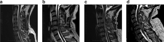
Magnetic resonance imaging (MRI) has become the gold standard for imaging neurological tissues including the spinal cord. The use of MRI for imaging in the acute management of patients with spinal cord injury has increased significantly. This paper used a vigorous literature review with Downs and Black scoring, followed by a Delphi vote on the main conclusions. MRI is strongly recommended for the prognostication of acute spinal cord injury. The sagittal T2 sequence was particularly found to be of value. Four prognostication patterns were found to be predictive of neurological outcome (normal, single-level edema, multi-level edema, and mixed hemorrhage and edema). It is recommended that MRI be used to direct clinical decision making. MRI has a role in clearance, the ruling out of injury, of the cervical spine in the obtunded patient only if there is abnormality of the neurological exam. Patients with cervical spinal cord injuries have an increased risk of vertebral artery injuries but the literature does not allow for recommendation of magnetic resonance angiography as part of the routine protocol. Finally, time repetition (TR) and time echo (TE) values used to evaluate patients with acute spinal cord injury vary significantly. All publications with MRI should specify the TR and TE values used.
Figures

References
-
- Andreoli C. Colaiacomo M.C. Rojas Beccaglia M. Di Biasi C. Casciani E. Gualdi G. MRI in the acute phase of spinal cord traumatic lesions: Relationship between MRI findings and neurological outcome. Radiol. Med. 2005;110:636–645. - PubMed
-
- Bondurant F.J. Cotler H.B. Kulkarni M.V. Mcardle C.B. Harris J.H., Jr. Acute spinal cord injury. A study using physical examination and magnetic resonance imaging. Spine (Phila Pa 1976) 1990;15:161–168. - PubMed
-
- Clark C.A. Werring D.J. Diffusion tensor imaging in spinal cord: Methods and applications – A review. NMR Biomed. 2002;15:578–586. - PubMed
-
- Como J.J. Thompson M.A. Anderson J.S. Shah R.R. Claridge J.A. Yowler C.J. Malangoni M.A. Is magnetic resonance imaging essential in clearing the cervical spine in obtunded patients with blunt trauma? J. Trauma. 2007;63:544–549. - PubMed
-
- Dai L. Jia L. Central cord injury complicating acute cervical disc herniation in trauma. Spine (Phila Pa 1976) 2000;25:331–335. discussion, 336. - PubMed
Publication types
MeSH terms
LinkOut - more resources
Full Text Sources
Other Literature Sources
Medical